41 trace a drop of blood through the heart diagram
Start studying Trace a drop of blood through the heart. Learn vocabulary, terms, and more with flashcards, games, and other study tools. Using a simple diagram to show the order in which blood flows through the heart, we will walk through the cardiac circulation pathway in 12 simple steps. As with every EZmed post, we have some simple tricks and charts that will help you remember the anatomy, physiology, and function of the right and left side of the heart.
Heart is a vital organ that you cannot live without. The function of heart is quite complex, but you can understand things better through the heart diagram labeled below. It provides information about different chambers of the heart and valves that help transfer blood from one part of your heart to another.
Trace a drop of blood through the heart diagram
SUMMARY1. Deoxygenated blood enters right atrium through Superior and Inferior Vena Cava2. Blood enters right ventricle through tricuspid ... In this interactive, you can label parts of the human heart. Drag and drop the text labels onto the boxes next to the diagram. Selecting or hovering over a box will highlight each area in the diagram. Right atrium: Segment of the heart that receives deoxygenated blood. Right atrium. The heart is made of four chambers which receive and pump blood. In the heart, the two circuits of the circulatory system (pulmonary and systemic circulation) converge. Before blood flows to the various parts of the body, it circulates in the heart and passes through the lungs.In this article, we will describe thepath of blood through the heart ...
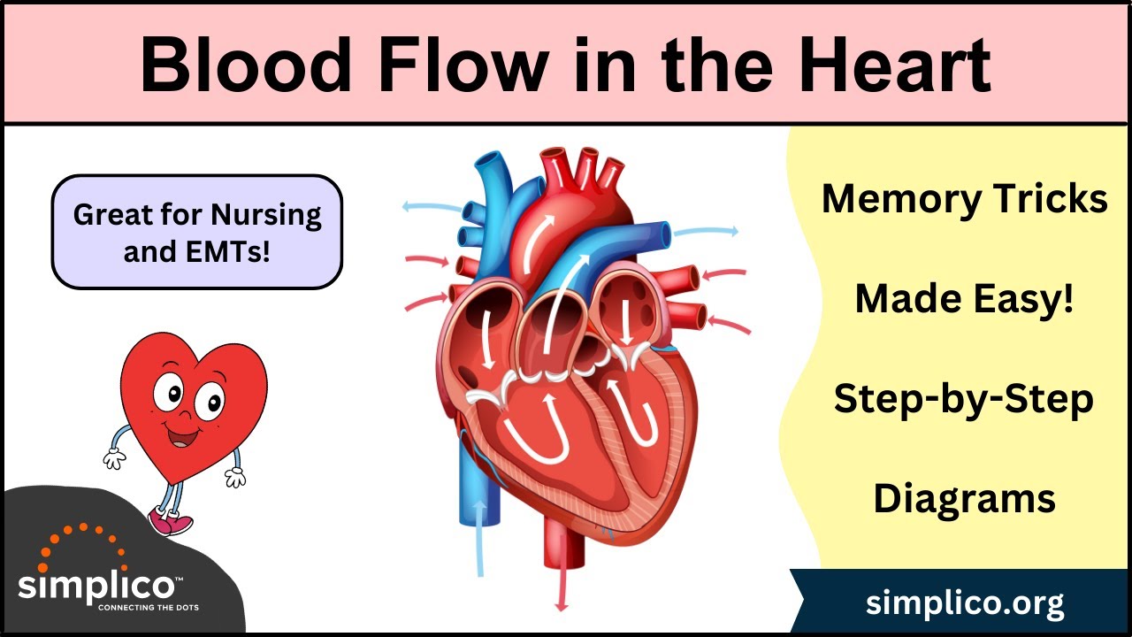
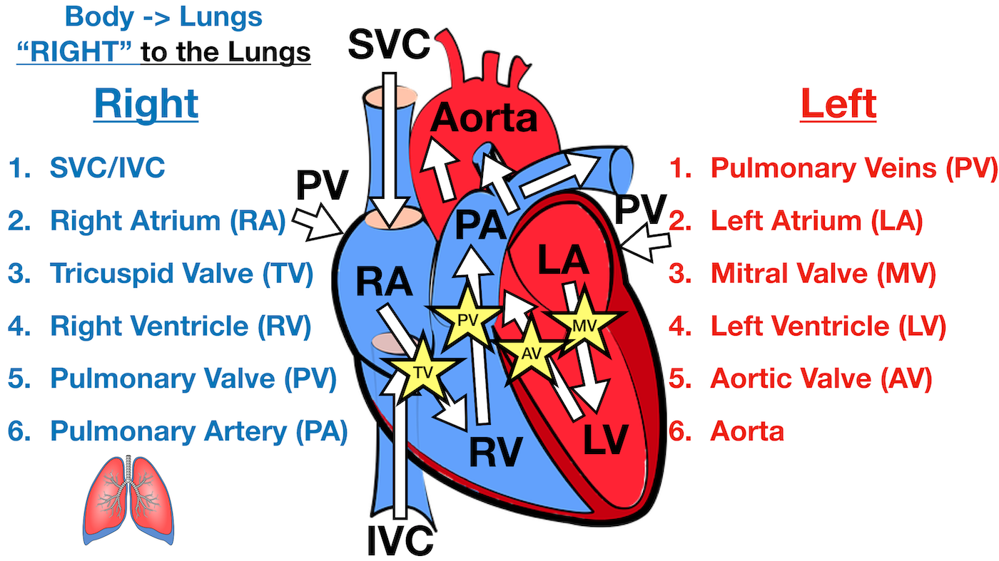
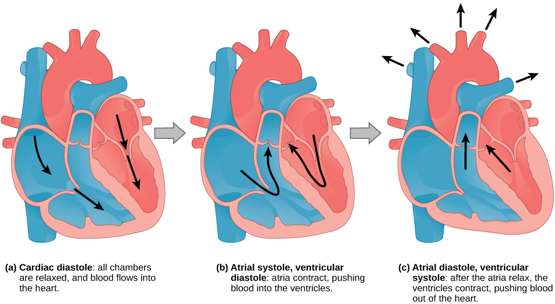
Trace a drop of blood through the heart diagram. The deoxygenated blood from the heart enters the lungs through the pulmonary valve as seen in the human heart diagram. This process is called pulmonary circulation. From the pulmonic valve, the blood travels to the pulmonary artery into the tiny capillary vessels of the lungs. The oxygen present in the tiny air sacs enters the blood through the ... Blood comes into the right atrium from the body, moves into the right ventricle and is pushed into the pulmonary arteries in the lungs. After ... The oxygen rich blood then returns to the fetus via the third vessel in the umbilical cord (umbilical vein). The oxygen rich blood that enters the fetus passes through the fetal liver and enters the right side of the heart. The oxygen rich blood goes through one of the two extra connections in the fetal heart that will close after the baby is born. Blood Flow Through the Heart. Blood flows through the heart in 12 easy steps. Always remember that it must flow through 6 areas on the right side and then 6 areas on the left side (this equals 12 steps). For better illustration, look at the picture below and note how the right and left side are separated.
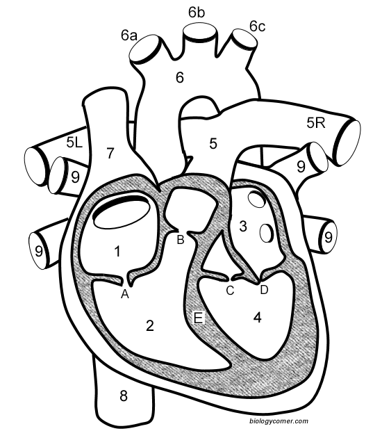
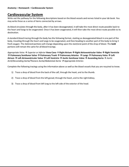
Trace a drop of blood through the heart from right atrium to the aorta. Locate and label the parts of a heart on a diagram. Compare the blood on the right side of the heart with that on the left side. Describe the components of blood.(red blood cells, white b.c., platelets and plasma) Trace a Drop of Blood Through the Body - Circulation Made Simple. The heart, a double-sided pump has the amazing ability to pump approximately 5 liters of blood through an average adult's body. Many people in medical terminology, anatomy and physiology, or other healthcare-related classes may find it helpful to review the pathways that blood ... The flow of blood through the heart should then be discussed. Arrows may also be placed on the diagram. Students can then "become"; a drop of blood, walk through the heart and trace the flow of blood. Oxygenated and deoxygenated blood may also be discussed. The diagram may then be left on the floor for several days and used for review. Pathway of Blood Through the Heart. In this educational lesson, we learn about the blood flow order through the human heart in 14 easy steps, from the superior and inferior vena cava to the atria and ventricles. Come also learn with us the heart's anatomy, including where deoxygenated and oxygenated blood flow, in the superior vena cava, inferior vena cava, atrium, ventricle, aorta ...
Blood flow through the heart. The heart is a muscular organ that pumps blood through the blood vessels of the circulatory system. Blood transports oxygen and nutrients to the body. It is also involved in the removal of metabolic wastes. This video describes how blood flows in and out of the heart. Blood Flow Through the Heart. Beginning with the superior and inferior vena cavae and the coronary sinus, the flowchart below summarizes the flow of blood through the heart, including all arteries, veins, and valves that are passed along the way. 1. Superior and inferior vena cavae and the coronary sinus. 2. The Right Ventricle. This is my Blood Path presentation. It will tell you how oxygenated blood from the toes travel through the body to the heart and lungs and back. The right ventricle then pumps the blood to the lungs so that the blood can pick up oxygen. First, the oxygen depleted blood starts in the toe, and travels to the heart through a vein. Figure 40.4 A. 1: View of the heart: This front view of the heart shows the direction of blood flow to and from the heart. Blood leaves the heart through the pulmonary artery and aorta, while blood enters the heart through the two venae cavae and pulmonary veins. The slow rate of travel through the capillary beds, which reach almost every cell ...
The blood then reaches the inferior vena cava, a major vein connected to the heart. Most of this blood is sent through the ductus venosus, also a shunt that passes highly oxygenated blood through the liver to the inferior vena cava and then to the right atrium of the heart. A small amount of this blood goes directly to the liver to give it the ...
The heart is an amazing organ. It starts beating about 22 days after conception and continuously pumps oxygenated red blood cells and nutrient-rich blood and other compounds like platelets throughout your body to sustain the life of your organs.; Its pumping power also pushes blood through organs like the lungs to remove waste products like CO2.; This fist-sized powerhouse beats (expands and ...

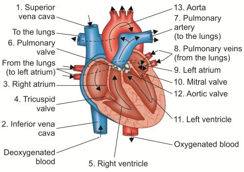
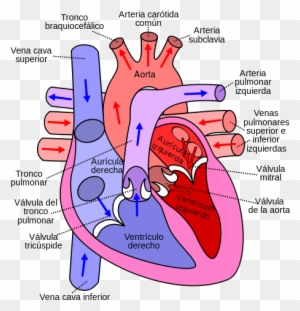
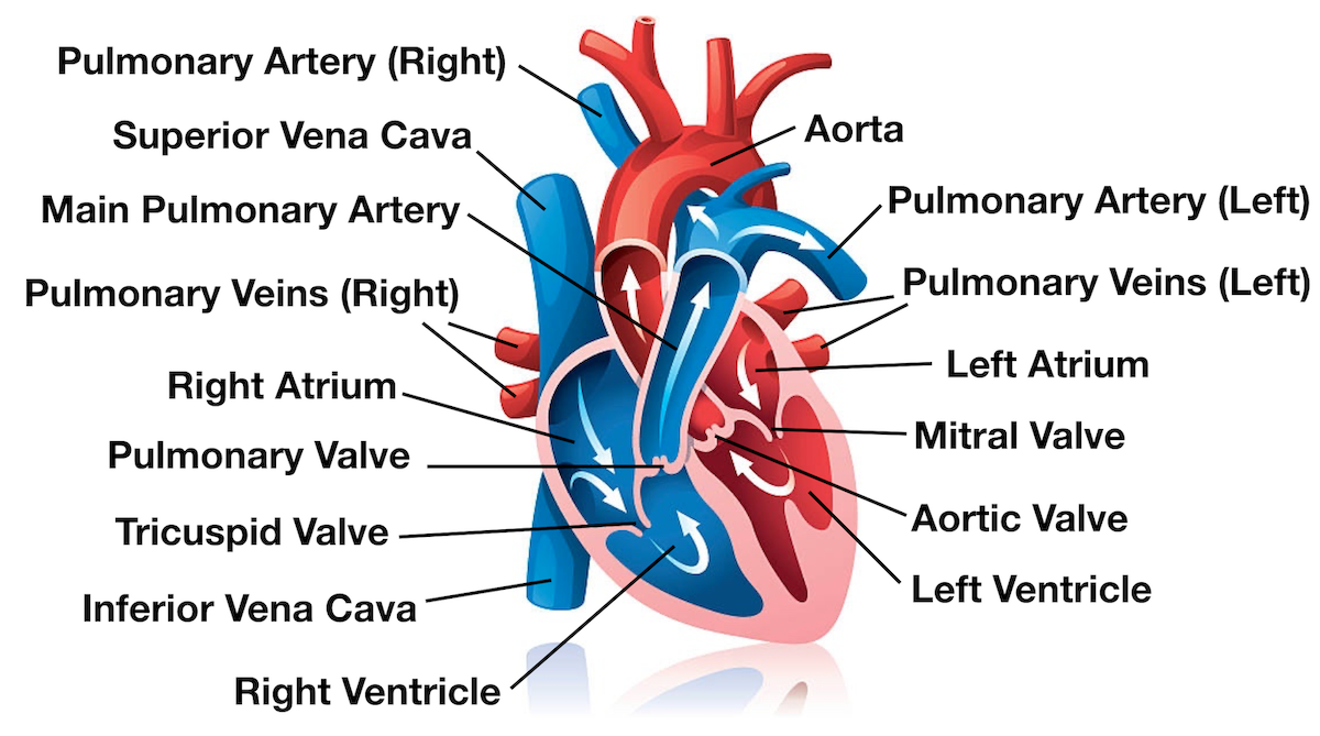
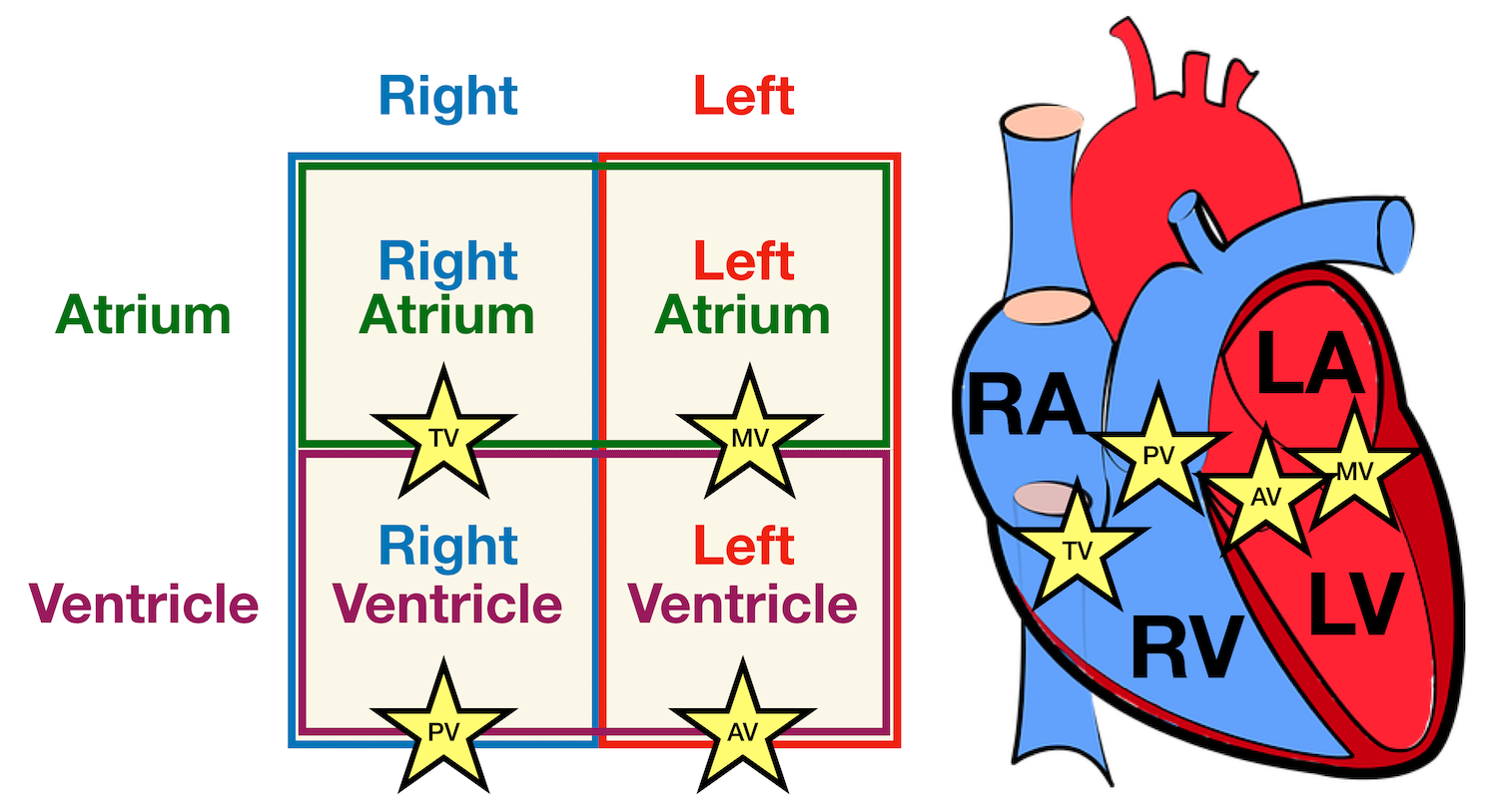
Pathway of blood through the heart 1. Blood enters the right atrium from the superior and inferior venae cavae, and the coronary sinus. 2. From right atrium, it goes through the tricuspid valve to the right ventricle. 3. From the right ventricle, it goes through the pulmonary semilunar valves to the pulmonary trunk 4.
/GettyImages-626974567-defc9220866c4e679c8d244dfbb997bb.jpg)
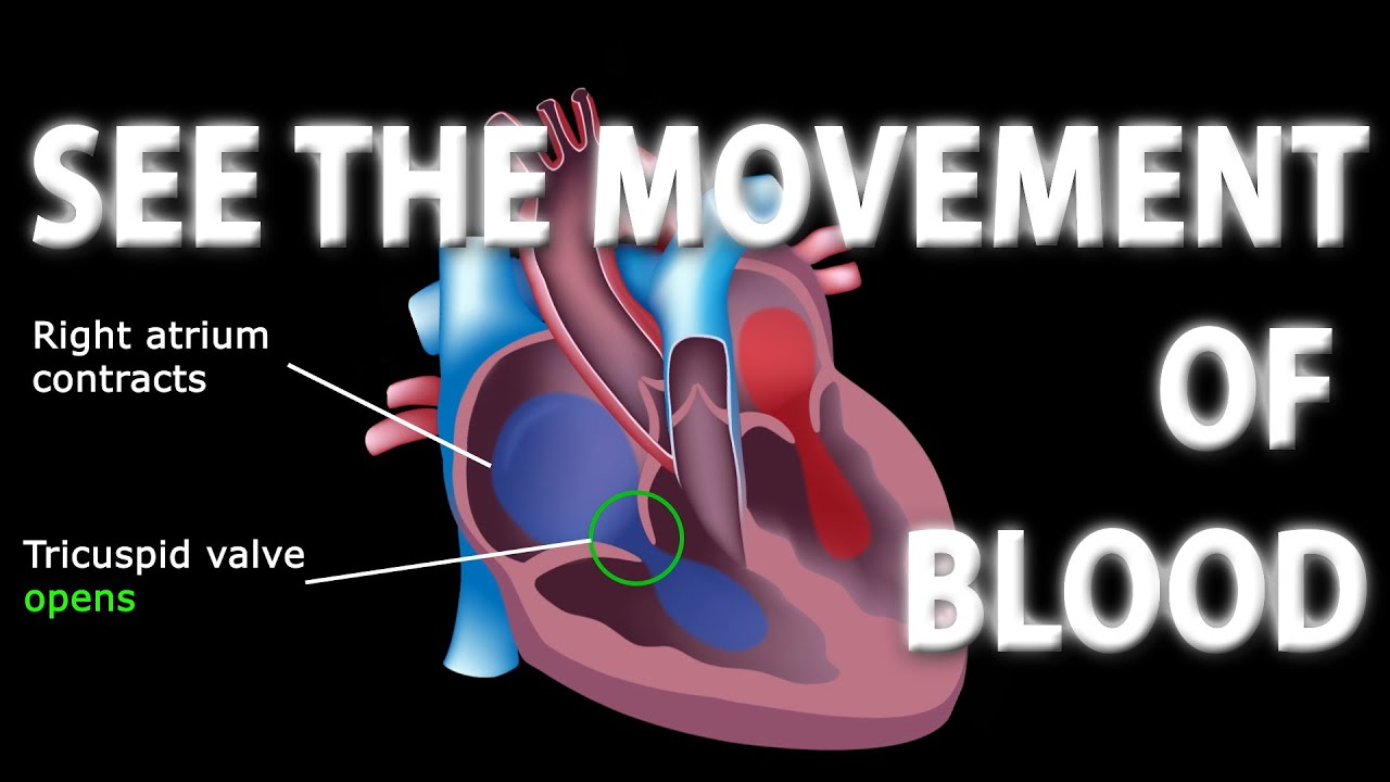
And once blood is in the right atrium, it's going to head down into the right ventricle. So this is the right ventricle, down here. This is the second chamber of the heart. And it gets there by passing through a valve. And this valve, and all valves in the heart, are basically there to keep blood moving in the right direction.

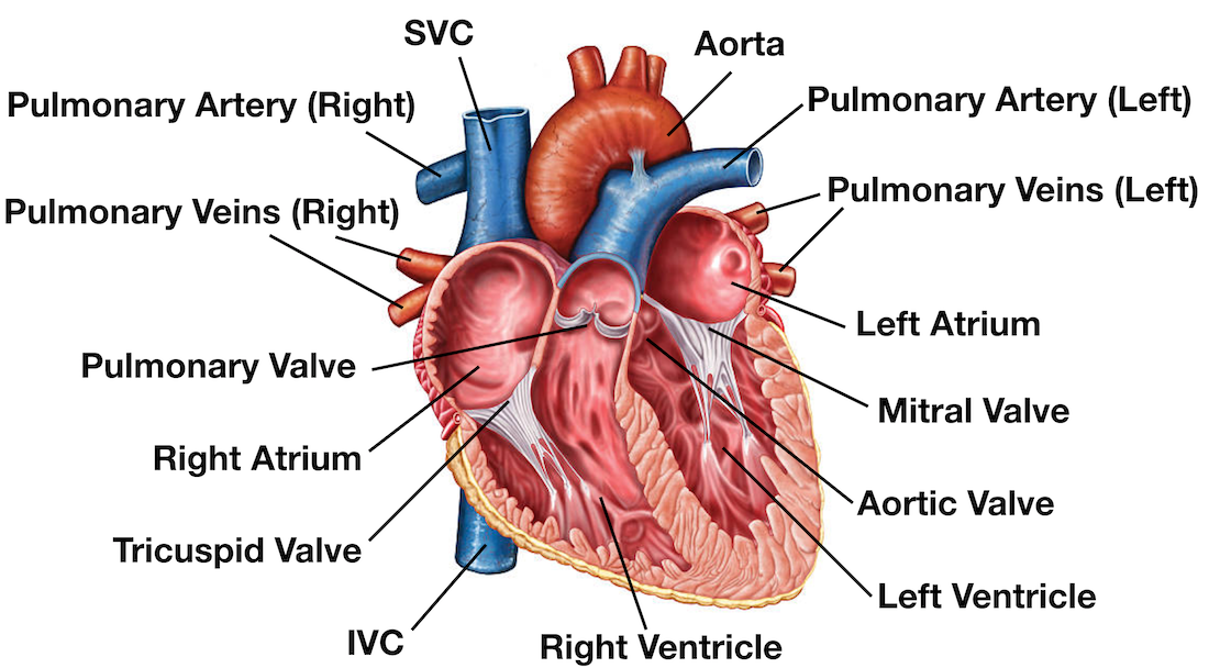
Arrows show the path of blood flow in the human heart. The blood enters the heart from the body through the superior vena cava and the inferior vena cava. Then the blood enters the right atrium chamber of the heart. The blood then moves through the tricuspid valve (shown as two white flaps) into the right ventricle chamber of the heart.
Trace the pathway of blood through the heart. Compare the pulmonary and systemic circuits. The heart has four hollow cavities, or chambers— two atria (a′tre-ah; singular atrium) and two ven-tricles (ven′tr˘ı-kulz). Each of these chambers is lined with endocardium, which helps blood flow smoothly through the heart. The superior atria are

In order to understand the disease processes (example: congenital heart defects) that affect the cardiac system, you must understand heart blood flow. This quiz will test your ability on how well you know the blood flow through the heart. Before taking the quiz, don't forget to watch the lecture on blood flow through the heart.

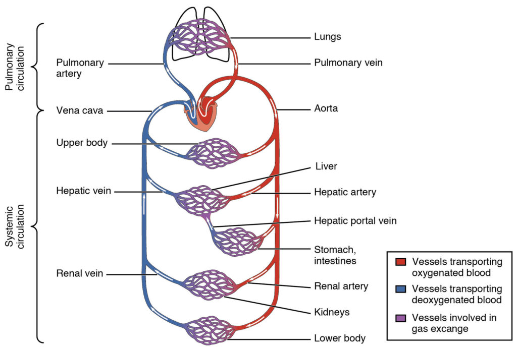
To trace a drop of blood through the entire circulatory system, we will start in the right atrium. Deoxygenated blood flows from the right atrium, through the tricuspid valve, to the right ventricle. From the right ventricle the blood flows through the pulmonary valve, to the pulmonary artery, leading to the lungs where the blood picks up oxygen. ...

Trace a drop of blood from the placenta into the fetal right atrium. ... There are 2 paths for blood through the fetal heart. Both are used. What are they? 1st path: RA-->tricuspid-->RV-->pulmonary valve-->pulmonary trunk-->ductus arteriosus-->aorta ... Trace the path through the fetal heart that uses the ductus arteriosus.
Blood enters the heart through two large veins, the inferior and superior vena cava, emptying oxygen-poor blood from the body into the right ...
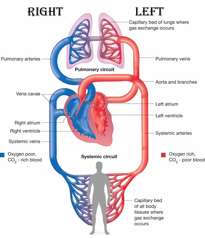
Circulatory Pathways. The blood vessels of the body are functionally divided into two distinctive circuits: pulmonary circuit and systemic circuit. The pump for the pulmonary circuit, which circulates blood through the lungs, is the right ventricle.The left ventricle is the pump for the systemic circuit, which provides the blood supply for the tissue cells of the body.
This video shows you how blood flows through the heart, out to the body, and back from the lungs. Having a strong sense of cardiac A&P is critical if you're ...
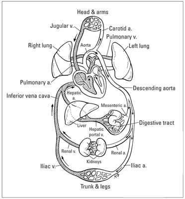
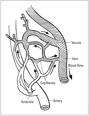
Objective 55: Trace the path of blood flow through the kidneys This diagram found on page 840 of our textbook was very helpful in learning the path of the blood flow through the kidneys. It is a very simple diagram, but it isn't confusing and it is very easy to remember.

Electrical impulses, controlled by the cardiac conduction system, make the heart muscle contract and relax, creating the rate and rhythm of your heartbeat. 1 Here are the steps of blood flow through the heart and lungs: The blood first enters the right atrium. The blood then flows through the tricuspid valve into the right ventricle.
Blood enters the heart through two large veins, the inferior and superior vena cava, emptying oxygen-poor blood from the body into the right ...
The heart is made of four chambers which receive and pump blood. In the heart, the two circuits of the circulatory system (pulmonary and systemic circulation) converge. Before blood flows to the various parts of the body, it circulates in the heart and passes through the lungs.In this article, we will describe thepath of blood through the heart ...

In this interactive, you can label parts of the human heart. Drag and drop the text labels onto the boxes next to the diagram. Selecting or hovering over a box will highlight each area in the diagram. Right atrium: Segment of the heart that receives deoxygenated blood. Right atrium.

SUMMARY1. Deoxygenated blood enters right atrium through Superior and Inferior Vena Cava2. Blood enters right ventricle through tricuspid ...


0 Response to "41 trace a drop of blood through the heart diagram"
Post a Comment