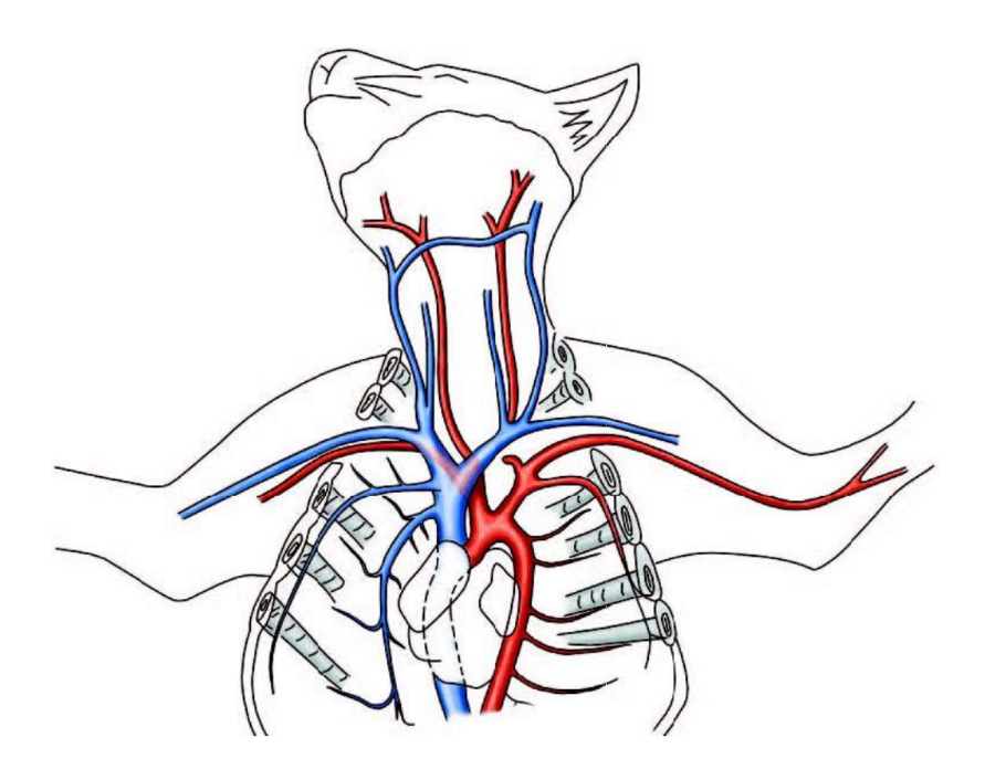
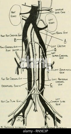
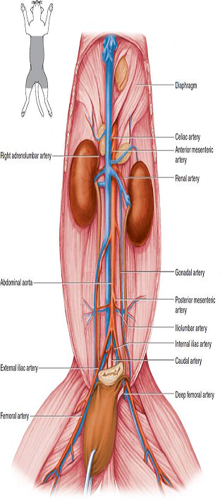
38 cat blood vessels diagram
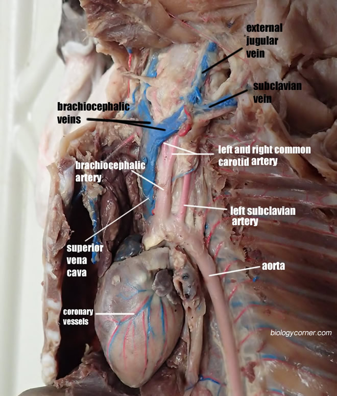
Cat Dissection - The Biology Corner Investigation: Blood Vessels above the Diaphragm. Double-injected cats are usually used to identify blood vessels. Arteries are injected with red latex, and veins are injected with blue latex. Blood vessels differ slightly in location from cat to cat. Carefully remove the fascia with blunt instruments to separate blood vessels from other ... Cat Dissection Blood Vessels | Biology Quiz - Quizizz Play this game to review Biology. To which vessel is #3 pointing? Preview this quiz on Quizizz. To which vessel is #3 pointing? Cat Dissection Blood Vessels DRAFT. 12th grade. 16 times. Biology. 66% average accuracy. 5 months ago. nbryant61. 0. Save. Edit. Edit. Cat Dissection Blood Vessels DRAFT. 5 months ago. by nbryant61. Played 16 times. 0 ...
Venous Blood Flow Diagram - blood flow restriction therapy ... Venous Blood Flow Diagram - 18 images - review visualize the system concepts images, the cardiovascular system basicmedical key, stomach artery homeopathy for all, pathophysiology of varicose veins plastic surgery key,

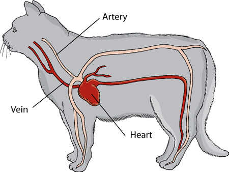
Cat blood vessels diagram
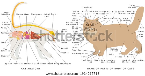
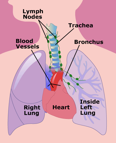
Cat and Dog Anatomy - VetMed The lymphatic system also works with the cardiovascular system to return fluids that escape from the blood vessels back into the blood stream. The digestive system ( cat ) ( dog ) includes the mouth, teeth, salivary glands, esophagus, stomach, intestine, pancreas, liver and gall bladder. PDF Exercise 32 Blood Vessels Eighth Edition deoxygenated blood Dissection of the Blood Vessels of the Cat • Read Dissection Exercise 4 (back of manual) before beginning your dissection. • We will dissect only the upper torso blood vessels (above the diaphragm) on the first dissection day, the lower torso blood vessels will be dissected on the second lab day. Heart Blood Flow | Simple Anatomy Diagram, Cardiac ... Diagram: Trick to remember the function of the left side of the heart is to pump oxygenated blood to the rest of the body - Blood that has "LEFT" to the lungs. Blood Flow of the Heart Review Let's now use the 2x2 table we made in the anatomy of the heart post, and this will give us another way to visualize the blood flow through the heart.
Cat blood vessels diagram. Eye Structure and Function in Cats - Cat Owners - Merck ... The orbit is a structure that is formed by several bones. The orbit also contains muscles, nerves, blood vessels, and the structures that produce and drain tears. The white of the eye is called the sclera. This is the relatively tough outer layer of the eye. It is covered by a thin membrane, called the conjunctiva, located near the front of the ... Blood Vessel: Overview, Anatomy, Types - Embibe Blood vessels carry the blood to deliver the nutrients to and remove wastes from our trillions of cells. Blood, heart and blood vessels together form the cardiovascular system. Blood vessels are divided into three types based on their structure and functions namely, arteries, veins, and capillaries. The study of blood vessels is often referred ... Cat Arteries and Veins Quiz - PurposeGames.com This is an online quiz called Cat Arteries and Veins. There is a printable worksheet available for download here so you can take the quiz with pen and paper. Your Skills & Rank. Total Points. 0. Get started! Today's Rank--0. Today 's Points. One of us! Game Points. 43. PDF Cat Dissection Arteries And Veins Labeling Quiz Dissection of the Blood Vessels of the Cat Dissection of April 10th, 2019 - Dissection of the Blood Vessels of the Cat Dissection Exercise 4 Objectives By the end of this lab you will be able to 1 Identify and label some of the most important blood vessels in the cat 2 Identify differences between the vascular system of the human and the cat 7 / 16
Differences Between Arteries and Veins - BYJUS Dec 14, 2021 · Arteries are the blood vessels that carry blood away from the heart, where it branches into even smaller vessels. Finally, the smallest arteries, called arterioles are further branched into small capillaries, where the exchange of all the nutrients, gases and other waste molecules are carried out.. Veins are the blood vessels present throughout the body. blood vessels in cat dissection - YouTube This video is part of a playlist on blood vessel anatomy & physiology at my youtube channel drjahn41. Feel free to review the other videos in the playlist at... Cat Skeleton Anatomy with Labeled Diagram » AnatomyLearner ... This simple guide might help you to get the basic anatomy of a cat skeleton. I always suggest you read the osteological features of any bones from a cat with a helpful diagram or model. The cat skeletal anatomy labeled diagrams that provide in this article might help you a lot. But if you need more cat anatomy labeled diagrams, please let me known. Blood Vessels • Types, Struture, Anatomy, & Functions Blood Vessels & Circulation. Tutorials and quizzes on the circulation of blood and the anatomy, structure, and physiology of blood vessels, using interactive animations and diagrams. Learn even faster with this blood vessel anatomy study guide. Learn anatomy faster and. remember everything you learn. Start Now.
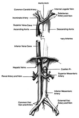
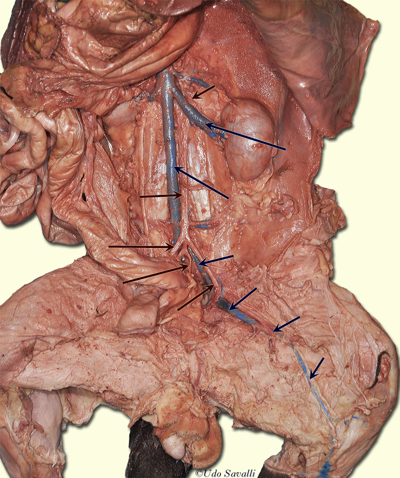
Pin by Lisa on A&P/Medicine/Pathology | Human heart ... Cat Circulatory System Lab Guide Answer Key Lab guide for the dissecting the circulatory system of the cat. It includes a list of the most common and easily identifiable vessels and sketches of the cat to label. Cat Arteries Quiz - PurposeGames.com This is an online quiz called Cat Arteries. There is a printable worksheet available for download here so you can take the quiz with pen and paper. From the quiz author. Cat Arteries Your Skills & Rank. Total Points. 0. Get started! Today's Rank--0. Today 's Points. One of us! Game Points. 26. cat blood vessels Flashcards - Quizlet What is its function. Descending (thoracic and abdominal) aorta. Major arteries that supplies thoracic and abdominal cavities with blood. both arteries seen. Brachiocephalic artery. sub divides into: right subclavian. R/L Common Carotid artery (in cat) one of two main branches off of Aorta in cat. Saddle Thrombus: Aortic Blood Clots in Cats The most common blockage point is in the lower abdomen where the aorta, the main blood vessel leaving the heart, forms two branches going to the back legs (**see Rough Diagram**). This spot is known as the saddle, and it is common for the blood clot to come to rest at the top of that point, leading to the term saddle thrombus.
Cat Eye Health - VetInfo The white part of the outer cat eye is called the sclera. Injury to the sclera is uncommon due to its strength. Occasionally, blood vessels become irritated and may burst, causing redness. Bruises to the sclera may indicate clotting issues or local injury. Yellowness of the sclera may indicate jaundice. Conjunctiva Should Resemble Gums in Color
Structure and Function of the Cardiovascular System in Cats A thrombus is a blood clot that develops within the heart or a blood vessel. An embolus is a blood clot that arises in one area of the circulatory system and is transported in the bloodstream to a distant site, where it becomes lodged in a blood vessel. The most common form of this disease in cats is the development of a blood clot in the left ...
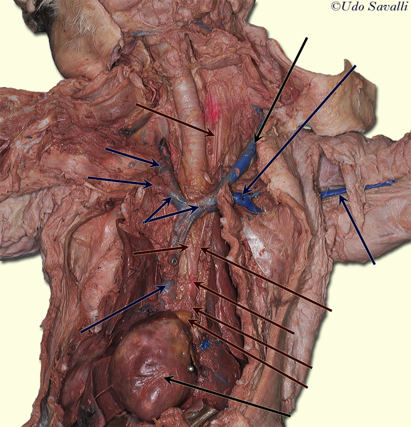
Cat Blood Vessels - Savalli Identify the blood vessels indicated by the arrows on the dissected cats. Arteries (red) Veins (blue) Aorta. Brachiocephalic Artery. Common Carotid Artery. Descending Aorta. External Iliac Artery. Femoral Artery.
Cat Vessels Image Gallery - The Biology Corner Cat Vessels Image Gallery. During this lab, students carefully teased the muscles and tissues away to reveal the arteries and veins. The goal was to identify each of the vessels on their lab guide and pass a lab test at the end of the activity where they are asked to locate the vessels.. After a Y incision is made, students carefully remove the pericardium and tissue surrounding the heart so ...
Cat and dog anatomy | Veterinary Teaching Hospital ... Dog Cat. Click the + to expand and see more information. The cardiovascular system includes the heart and blood vessels. The cardiovascular system performs the function of pumping and carrying blood to the rest of the body. The blood contains nutrients and oxygen to provide energy to allow the cells of the body to perform work.
Arteries of the Hindlimb - Anatomy & Physiology - WikiVet Introduction. The abdominal aorta terminates by branching into the external iliac arteries and the internal iliac arteries.It is these arteries that supply the hindlimb and pelvis. (Note: Although the information below is based around the anatomy of the canine hindlimb, it is essentially the anatomy of the arteries in domestic species.
cat blood vessels Flashcards | Quizlet Not all inclusive, check your study sheets for complete list. Learn with flashcards, games, and more — for free.
Cat Circulation - Murray State University Cat Circulation. Click on a number below to see blood vessels in the area specified.
Animal Vascular Anatomy - Long Beach Animal Hospital The aorta is one of many blood vessels in the chest This radiograph of a cat's chest shows the aorta as it leaves the heart in the same way as the picture above. It is the grayish linear object with arrows on top. The arrows show the direction of blood flow to the back of the body.
Anatomy and Physiology - Dr. Scott Croes' Website Labeling Exercises for Blood Vessels Labeling Exercises for the Cat Blood Vessels and Sheep Heart Heart Diagram for Coloring Animation: Autorhythmic Cells Cardiac Action Potential Animation: Complete Review of the Heart Animation: Conducting System of the Heart Animation: The Cardiac Cycle Animation: The Cardiac Cycle Part II
Your Eyes (for Kids) - Nemours KidsHealth These are blood vessels, the tiny tubes that deliver blood, to the sclera. The cornea (say: KOR-nee-uh), a transparent dome, sits in front of the colored part of the eye. The cornea helps the eye focus as light makes its way through.
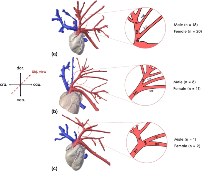
PDF Respiratory System - Stevenson High School In the cat, the aortic arch gives off two large vessels, the right brachiocephalic arteryand the left subclavian artery. The right brachiocephalic arteryhas three major branches, the right subclavian arteryand the right and left common carotid arteries. (Note that in humans, the left common carotid artery is a direct branch off the aortic arch.)
Circulatory System - Pennsylvania State University The following links will allow you to access real photographs of the cat circulatory system. The purpose of these pages is to quiz your knowledge on the structures of the circulatory system. Please try to answer all structures (or guess) before you look at the answers! Choose one of the following categories: Thoracic Cavity Anatomy
Structure and Functions of Human Eye with labelled Diagram Jun 24, 2021 · Human Eye Diagram: Contrary to popular belief, the eyes are not perfectly spherical; instead, it is made up of two separate segments fused together. Explore: Facts About The Eye To understand more in detail about our eye and how our eye functions, we need to look into the structure of the human eye.
Heart Blood Flow | Simple Anatomy Diagram, Cardiac ... Diagram: Trick to remember the function of the left side of the heart is to pump oxygenated blood to the rest of the body - Blood that has "LEFT" to the lungs. Blood Flow of the Heart Review Let's now use the 2x2 table we made in the anatomy of the heart post, and this will give us another way to visualize the blood flow through the heart.
PDF Exercise 32 Blood Vessels Eighth Edition deoxygenated blood Dissection of the Blood Vessels of the Cat • Read Dissection Exercise 4 (back of manual) before beginning your dissection. • We will dissect only the upper torso blood vessels (above the diaphragm) on the first dissection day, the lower torso blood vessels will be dissected on the second lab day.
Cat and Dog Anatomy - VetMed The lymphatic system also works with the cardiovascular system to return fluids that escape from the blood vessels back into the blood stream. The digestive system ( cat ) ( dog ) includes the mouth, teeth, salivary glands, esophagus, stomach, intestine, pancreas, liver and gall bladder.















0 Response to "38 cat blood vessels diagram"
Post a Comment