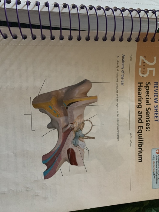
39 identify all indicated structures and ear regions in the following diagram
Labeled Sarcomere Diagram Dodge Durango Wiring Diagram. Start studying UNIT 5: Label the parts of the Sarcomere. Learn vocabulary, terms, and more with flashcards, games, and other study tools. Draw and label a diagram to show the structure of a sarcomere, including Z lines, actin filaments, myosin filaments with heads, and the resultant light and dark bands. EX 25 Special Senses: hearing and Equilibrium - Quizlet Identify all indicated structures and ear regions in the following diagram refer to diagram Sacs found within the vestibule Saccule, utricle contains the spiral organ Cochlear duct Sites of the maculae Saccule, utricle Positioned in all spatial planes Semicircular ducts Hair cells of spiral organ rest on this membrane Basilar membrane
PDF Body regions, Major body Cavities - Sinoe Medical Association Body regions Right or left based on the person being viewed. ... • which houses all other internal body organs 1). Thoracic: ... Middle Ear Cavities. 5). Synovial Cavities. Membranes

Identify all indicated structures and ear regions in the following diagram
Solved Review Sheet: Special Senses: Hearing and ... Experts are tested by Chegg as specialists in their subject area. We review their content and use your feedback to keep the quality high. Transcribed image text: Review Sheet: Special Senses: Hearing and Equilibrium 2. Identify all indicated structures and ear regions in the following diagram. or. blog.bestwriters.org › 2020/09/23 › data-structuresData Structures And Algorithm Analysis - Best Writers Sep 23, 2020 · Question is in the file, just two questions but need to be down by 10pm today. The book is uploaded. thanks for helping. 1. In the probability review in class, we showed an easier way to compute the Expected value (average) of the sum of two dice, by using the Linearity of Expectations. (a) Show […] DOC 142 Anatomy & Physiology Coloring Workbook Using the key choice terms given in Exercise 3, identify the structures indicated by leader lines on the diagram of the eye in Figure 8-2. Select different colors for all structures provided with a color-coding circle and then use them to color the coding circles and corresponding structures in the figure. 6.
Identify all indicated structures and ear regions in the following diagram. › science › articleTethering Piezo channels to the actin cytoskeleton for ... Feb 08, 2022 · (A) Diagram showing the central pore module of Piezo1 containing the indicated domains and the topological representation of E-cadherin containing the N-terminal ectodomain (ED), the single transmembrane region (TM), and the intracellular C-terminal tail (CT). (B–F) Representative western blots under the indicated conditions. Senses Quiz Flashcards | Quizlet Correctly identify the following structures of the cochlea. Place the following labels in order indicating the passage of sound waves and their conversion to fluid waves through the ear and hearing apparatus. 1. auricle 2. auditory canal 3. tympanic membrane 4. malleus 5. incus 6. stapes 7. oval window 8. scala vestibuli 9. cochlear duct PDF 2 the Anatomy and Physiology of The Ear and Hearing The ear canal is about 4 centimetres long and consists of an outer and inner part. The outer portion is lined with hairy skin containing sweat glands and oily sebaceous glands which together form ear wax. Hairs grow in the outer part of the ear canal and they and the wax serve as a protective barrier and a disinfectant. Label Parts of the Human Ear - University of Dayton Parts of the Ear. Select the correct label for each part of the ear. Click on the Score button to see how you did. Incorrect answers will be marked in red.
Human Ear: Structure and Functions (With Diagram) ADVERTISEMENTS: In this article we will discuss about the structure and functions of human ear. Structure of Ear: Each ear consists of three portions: (i) External ear, ADVERTISEMENTS: (ii) Middle ear and (iii) Internal ear. 1. External Ear: It comprises a pinna, external auditory meatus (canal) & tympanic membrane. (i) Pinna: ADVERTISEMENTS: The pinna is […] [Solved] 386 Review Sheet 25 2. Identify the structures of ... A. Middle ear - it mainly consists of 3 bones, which help in amplification and transmission of sound wave from tympanic membrane to cochlea. The 3 bones are - malleus, incus and stapes B. Inner ear It consists of cochlea - it contains receptor for hearting. Semicircular canals and vestibule - help in balance. Step-by-step explanation Using the above referenced figures of the anatomy of a ... Using the above-referenced diagram of the anatomical distribution of sympathetic postganglionic fibers, identify the specified labeled structure (s) in each of the following questions. 20) Identify the structure (s) indicated by Label I. A) Spinal cord B) Medulla oblongata C) Hypothalamus D) Pons E) Midbrain › cms › lib2Fetal Pig Dissection Lab - Humble Independent School District Be sure to be able to identify both male and female pigs and their reproductive structures. Locate all three openings (urethral opening, vaginal orifice, and anus) on the female pig. The urethral opening excretes urine and the vaginal orifice is the opening of the birth canal. In males, the urogenital structures consist
Practical worksheet 5 (JAN 2021) The Sensory System ... Identify all indicated structures and ear regions that are provided with leader lines or brackets in the following diagram. 5. Match the membranous labyrinth structures in the listed key with the descriptive statements below. Ch.25 Special senses: hearing and equilibrium - Quizlet STRUCTURE COMPOSING THE EXTERNAL EAR PINNA (AURICLE), EXTERNAL AUDITORY CANAL, TYMPANIC MEMBRANE STRUCTURES COMPOSING THE INTERNAL EAR COCHLEA, SEMICIRCULAR CANALS, VESTIBULE COLLECTIVELY CALLED THE OSSICLES INCUS (ANVIL), MALLEUS (HAMMER), STAPES (STIRRUP) INVOLVED IN EQUALIZING THE PRESSURE IN THE MIDDLE EAR WITH ATMOSPHERIC PRESSURE The Human Ear - Structure, Functions and its Parts The ear is a sensitive organ of the human body. It is mainly concerned with detecting, transmitting and transducing sound. Maintaining a sense of balance is another important function performed by the human ear. Let us have an overview of the structure and functions of the human ear. Structure of Ear. The human ear consists of three parts ... Solved: Chapter E17 Problem 21E Solution - Chegg There are three parts of the ear, namely the external, internal, and the middle ear. The external ear comprises of the auditory canal (external) and the pinna. The middle one comprises of the ossicles, and eustachian tube. The external, and the middle ear is divided by the tympanic membrane.
Ear Anatomy: Pictures Flashcards - Quizlet Identify I: Semicircular Canals. Identify J: pinna/auricle. Identify A: Cochlea. Identify I: a coiled, bony, fluid-filled tube in the inner ear through which sound waves trigger nerve impulses. External Auditory meatus.
Chapter 11. Fetal Pig Dissection - Anatomy and Physiology ... The following words will be used to help identify the location of structures. Anterior refers to the head end. If a structure is anterior to another then it is closer to the head. Posterior refers to the tail end. Dorsal refers to the back side. The pig in the first photograph below is laying on its dorsal side. Ventral is the belly side.
1.4 Anatomical Terminology - Anatomy & Physiology Figure 1.4.1 - Regions of the Human Body: The human body is shown in anatomical position in an (a) anterior view and a (b) posterior view. The regions of the body are labeled in boldface. A body that is lying down is described as either prone or supine.
Human Ear Anatomy - Parts of Ear Structure, Diagram and ... Human ear. The ear is divided into three anatomical regions: the external ear, the middle ear, and the internal ear (Figure 2). The external ear is the visible portion of the ear, and it collects and directs sound waves to the eardrum. The middle ear is a chamber located within the petrous portion of the temporal bone.
› pmc › articlesMobile Genetic Elements Associated with Antimicrobial ... Aug 01, 2018 · Insertion points for resistance regions common to plasmids of the same type are also indicated, in some cases, by labeled vertical arrows. C backbones are represented by a single line, with differences (presence/absence of ARI-A, orf1832 versus orf1847 , rhs1 versus rhs2 , and presence/absence of i1 and i2) shown above (C 1 ) and below (C 2 ).
Mobile Genetic Elements Associated with Antimicrobial ... 01.08.2018 · It is not possible to cover all MGE involved in resistance in ... Diagrams were drawn based on sequences from the following INSDC accession numbers: IS 1247, AJ971344; ISKpn23, KP689347; ISEnca1, AY939911 (end of TPU found by alignment with Staphylococcus plasmids, e.g., pSTE1 [accession number HE662694]); and ISEcp1, FJ621588. (E) Different …
› nervous-system-sense-organsICSE Solutions for Class 10 Biology – The ... - A Plus Topper Dec 04, 2019 · Question 13: Study the following diagram carefully and then answer the questions that follow. The diagram is depicting a defect of the human eye: (i) Identify the defect shown in the diagram. (ii) Give two possible reasons for the above defect. Answer: (i) Far sightedness or hypermetropia. (ii) (a) Lens is flattened or less convex, (b) Eyeball ...
Structure and Functions of the Ear Explicated With ... Outer ear is divided into the pinna and the external auditory meatus. The pinna, also known as the auricle is the external ear part that is located and seen on each side of our head. It is made up of cartilage and soft tissue. This helps in maintaining a particular ear shape and remains pliable.
The Skull | Anatomy and Physiology I - Lumen Learning The cranium (skull) is the skeletal structure of the head that supports the face and protects the brain.It is subdivided into the facial bones and the brain case, or cranial vault (Figure 1).The facial bones underlie the facial structures, form the nasal cavity, enclose the eyeballs, and support the teeth of the upper and lower jaws.
Genome-Wide Analysis of the ERF Gene Family ... - OUP Academic To identify the ERF family genes in Arabidopsis, ... this same alignment indicated that the C-terminal regions of the AP2/ERF domains of seven proteins (AtERF#116–AtERF#122) possess a very low homology to the consensus sequence (Fig. 1. Figure 1. Open in new tab Download slide. Alignment of the AP2/ERF domains from Arabidopsis ERF proteins. Black and light gray …
fluidcontainedwithinthebonylabyrinthandbathingthe ... 4. Identify all indicated structures and ear regions in the following diagram. 5. Match the membranous labyrinth structures listed in column B with the descriptive statements in column A. Some terms are used more than once.
PDF Practice Quiz Tissues - Portland Community College Identify the structure indicated. Compact Bone (Osseous Tissue) Central Canal. Identify the tissue type and a location where it is found. Elastic Cartilage •External Ear •Epiglottis. Identify the structure indicated. Simple Squamous Epithelium: Basement Membrane. Identify the tissue type and its function.
Anatomy of the Ear | Inner Ear | Middle Ear | Outer Ear The Outer Ear The outer ear includes: auricle (cartilage covered by skin placed on opposite sides of the head) auditory canal (also called the ear canal) eardrum outer layer (also called the tympanic membrane) The outer part of the ear collects sound. Sound travels through the auricle and the auditory canal, a short tube that ends at the eardrum.
academic.oup.com › plphys › articleGenome-Wide Analysis of the ERF Gene Family in ... - OUP Academic Jan 11, 2006 · The phylogenetic tree, location of the intron (arrowhead), and a schematic diagram of the protein structures of every group, I to VI, VI to L, VII to X, and Xb-L, are shown in A to L, respectively. Each colored box represents the AP2/ERF domain and conserved motifs, as indicated below the tree.
Study Parts of the Ear (Anatomy) Flashcards | Quizlet Study with Flashcards again. 1/34. Created by. Meghan_Brooks7. Identify the part of the ear that is shaded blue. Images derived from an image by Chittka L, Brockmann's under a Creative Commons license (but not endorsed by the author). The length of the auditory canal is exaggerated for viewing purposes.
DOC 142 Anatomy & Physiology Coloring Workbook Using the key choice terms given in Exercise 3, identify the structures indicated by leader lines on the diagram of the eye in Figure 8-2. Select different colors for all structures provided with a color-coding circle and then use them to color the coding circles and corresponding structures in the figure. 6.
blog.bestwriters.org › 2020/09/23 › data-structuresData Structures And Algorithm Analysis - Best Writers Sep 23, 2020 · Question is in the file, just two questions but need to be down by 10pm today. The book is uploaded. thanks for helping. 1. In the probability review in class, we showed an easier way to compute the Expected value (average) of the sum of two dice, by using the Linearity of Expectations. (a) Show […]
Solved Review Sheet: Special Senses: Hearing and ... Experts are tested by Chegg as specialists in their subject area. We review their content and use your feedback to keep the quality high. Transcribed image text: Review Sheet: Special Senses: Hearing and Equilibrium 2. Identify all indicated structures and ear regions in the following diagram. or.

0 Response to "39 identify all indicated structures and ear regions in the following diagram"
Post a Comment