38 Ventral Body Cavity Diagram
Major Body Cavities, Their Subdivisions And Membranes. - Earth's Lab The ventral body cavity organs are supported as well as protected by serosae (singular, serosa), or serous membranes. The serous membranes are thin layers of tissue that line the body cavity and protect the internal organs. Serous membranes have a superficial parietal layer which lines the cavity... BLANK ventral body cavity diagram - | Course Hero View Homework Help - BLANK ventral body cavity diagram from BIO 208 at California State University, Long Beach.
40+ Ventral body cavity PowerPoint (PPT) Presentations... - SlideServe SlideServe has a very huge collection of Ventral body cavity PowerPoint presentations. You can view or download Ventral body cavity presentations for your school assignment or business presentation. Browse for the presentations on every topic that you want.

Ventral body cavity diagram
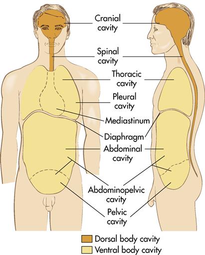
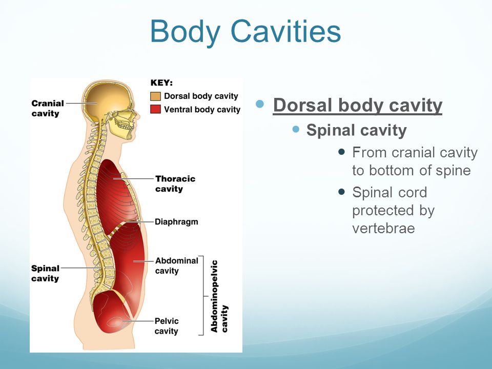
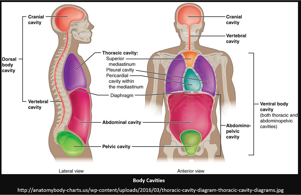
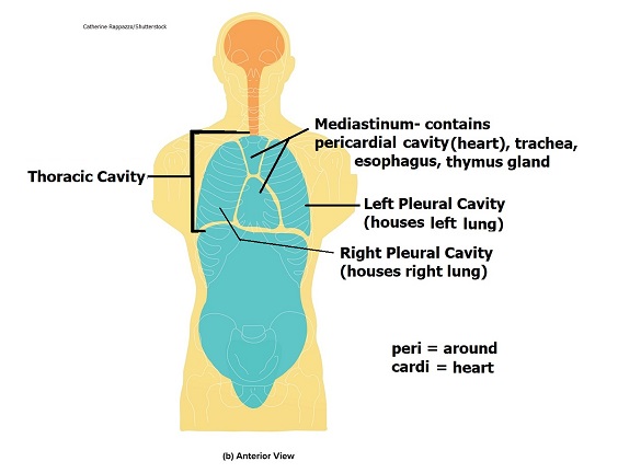
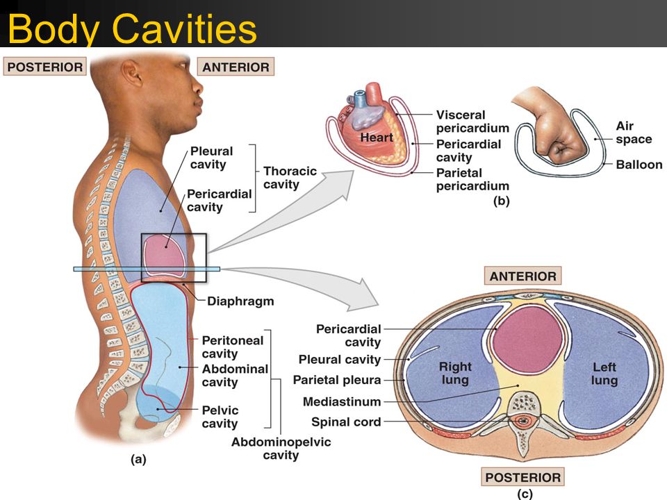
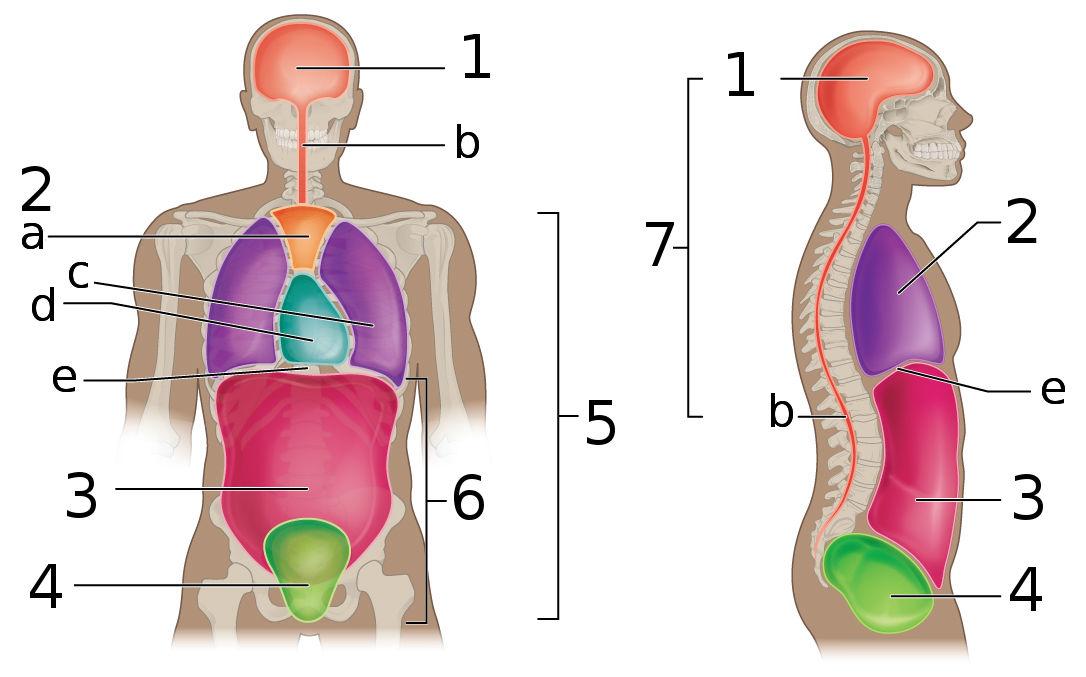
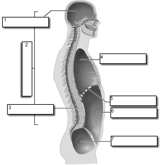
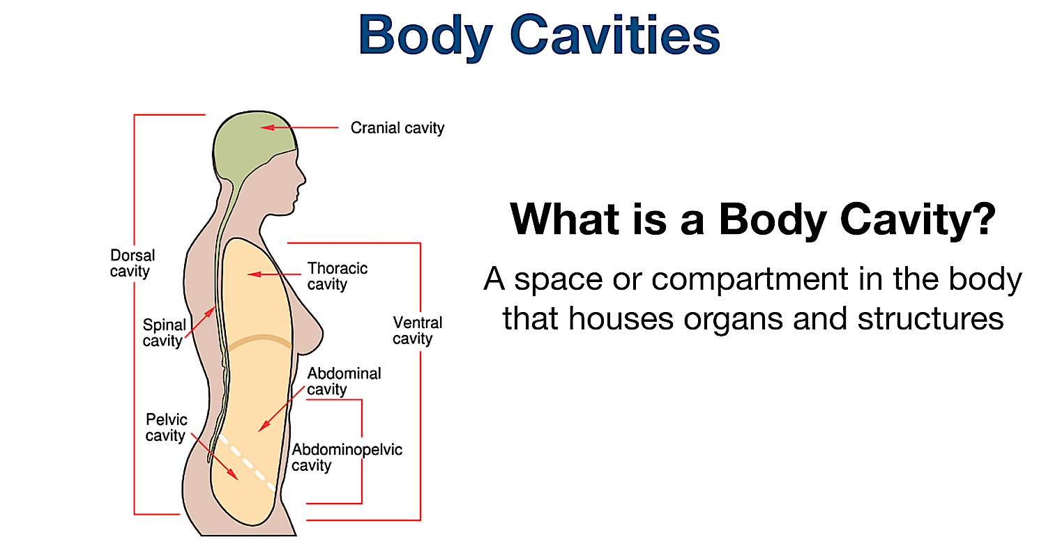
Body Cavities Labeling Shows the body cavities from a front view and a lateral view, practice naming the cavity by filling in the boxes. Front View: 1. Cranial Cavity 2. Vertebral Canal 3. Mediastinum 4. Pleural Cavity 5. Pericardial Cavity 6. Diaphragm 7. Abdominal Cavity 8. Pelvic Cavity 9. Abdominopelvic Cavity 10. Body Cavities Ventral body cavity (thoracic and abdominopelvic cavities) Ventral Body Cavity • Houses internal organs (viscera) • Two subdivisions (separated by diaphragm) - Thoracic cavity - Abdominopelvic cavity © 2013 Pearson Education, Inc. Ventral Body Cavity • Thoracic cavity subdivisions... Body cavity - New World Encyclopedia In zoology, body cavity generally refers to the space, or cavity, located between an animal's outer covering (epidermis) and the outer lining of the gut cavity—a fluid-filled space where internal organs develop.
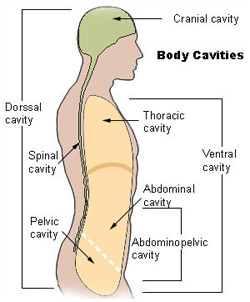
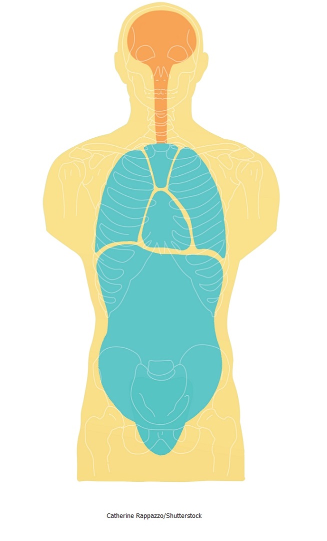
Ventral body cavity diagram. Ventral Cavity - Definition and Function | Biology Dictionary Like all body cavities, the ventral cavity has several important functions relating to the organs housed within in. First and foremost, the cavity protects the organs inside from shock damage as the organism moves through the world. The space and fluid around the organs insures that any impacts incurred by... Body cavity Body cavity, Dorsal mesentery → Double layers - Coggle Diagram: Body cavity (Dorsal & Ventral tube, End of the 4th week: Closing of the ventral body wall, Serous membranes, Formation body cavity ( End 3rd week)), Dorsal mesentery...Create your own diagrams like this for free with Coggle. Body Cavities and Organs with Labeled Diagram - The Major and... You will also find the body cavities and organs labeled diagram so that you may identify them so quickly from the actual sample. The basal lamina of the ventral nasal concha curves medially and ventrally into the cavity. The conchae divide the nasal cavity into four primary passages (known as... Diagram of Dorsal and ventral body cavities | Quizlet Abdominal Cavity. contains digestive viscera. Pelvic Cavity. contains urinary bladder, reproductive organs, and rectum.
Dorsal and Ventral Body Cavities - YouTube Based on ANAT113 from Centennial College, this channel is designed to help students understand the tricky topics of Anatomy and Physiology.Share your... Body cavity - New World Encyclopedia In zoology, body cavity generally refers to the space, or cavity, located between an animal's outer covering (epidermis) and the outer lining of the gut cavity—a fluid-filled space where internal organs develop. Body Cavities Ventral body cavity (thoracic and abdominopelvic cavities) Ventral Body Cavity • Houses internal organs (viscera) • Two subdivisions (separated by diaphragm) - Thoracic cavity - Abdominopelvic cavity © 2013 Pearson Education, Inc. Ventral Body Cavity • Thoracic cavity subdivisions... Body Cavities Labeling Shows the body cavities from a front view and a lateral view, practice naming the cavity by filling in the boxes. Front View: 1. Cranial Cavity 2. Vertebral Canal 3. Mediastinum 4. Pleural Cavity 5. Pericardial Cavity 6. Diaphragm 7. Abdominal Cavity 8. Pelvic Cavity 9. Abdominopelvic Cavity 10.
Body cavity - Wikipedia

Ventral Body Cavity | Subdivisions, Organs, & Diagram - Video ...

Body Cavities and Membranes – Anatomy and Physiology Notes
1. Introduction to anatomy and physiology | Nurse Key

Body Cavities and Membranes – Anatomy and Physiology Notes
Body Cavities. Dorsal body cavity Cranial cavity Space inside ...

Dorsal and ventral body cavities Flashcards | Quizlet
Body Cavities and Membranes - SCIENTIST CINDY

Chapter 1: Part 6: Body Planes & Body Cavities - ppt download
Body Cavities and Membranes – Anatomy and Physiology Notes
![Anatomical Terminology | Anatomy and Physiology I [ARCHIVED]](https://s3-us-west-2.amazonaws.com/courses-images-archive-read-only/wp-content/uploads/sites/198/2014/10/20082356/107_Regions_of_Human_Body_new.jpg)
Anatomical Terminology | Anatomy and Physiology I [ARCHIVED]
Aim: How can we identify and describe the human body cavities ...

Ch.1 ventral & dorsal cavities Diagram | Quizlet

Body Cavities Quiz

The ventral body cavity is subdivided into the ______ and the ...

Chapter 1H Body Cavities
File:Body Cavities labeled.png - Wikimedia Commons

Representation of the ventral body cavity, made up of the ...
SEER Training: Anatomical Terminology

Body cavities

VENTRAL cavity (ventre = stomach) and DORSAL cavity (dorsal ...

Dorsal and Ventral Body Cavities · Open Educational Resource ...
Biology 2404 Human A&P Basics
Body Cavities Labeling

Preparing for A&P: Basic Science and Biology, 1st Edition ...
Body Cavities and Membranes Quiz Anatomy and Physiology
Directional Terms, Planes, Sections, Body Cavities Flashcards ...

Body Cavities Flashcards | Quizlet

Body anatomy, Thoracic cavity, Medical dictionary
Shutterstock - PuzzlePix
Welcome to Anatomy and Physiology
Introduction to Anatomy, Chapter 1

Ventral body cavity - wikidoc
Bio 103 Introduction - Student Handout.pptx
Anatomical Terminology
Body Cavities Study Guide: Labeled Organs and Membranes

The Human Body Cavities | Human Anatomy and Physiology Lab ...

Ventral Body Cavity: Definition, Subdivisions & Organs Video
0 Response to "38 Ventral Body Cavity Diagram"
Post a Comment