37 Inner Ear Crystals Diagram
Ear Crystals - Undizzy Me Ear crystals and craniosacral vertigo are two different types of vertigo with different causes and different Vertigo caused by ear crystals is called Benign Paroxysmal Positional Vertigo or BPPV. The crystals are actually debris that have migrated from another part of the inner ear where they do... Dizziness inner ear crystals Dizziness inner ear crystals,definition type 2 diabetes mellitus,1200 calorie diet planner - Reviews. Bacterial or viral infection sometimes in association with allergy leads to an accumulation of fluid in the middle ear space behind the ear drum (see ear diagram).
Skeletal System – Labeled Diagrams of the Human Skeleton 29.7.2020 · Found in a small cavity inside of the temporal bone, they serve to transmit and amplify sound from the eardrum to the inner ear. Vertebrae. Twenty-six vertebrae form the vertebral column of the human body. They are named by region: Cervical (neck) - 7 vertebrae; Thoracic (chest) - 12 vertebrae; Lumbar (lower back) - 5 vertebrae; Sacrum - 1 vertebra

Inner ear crystals diagram
The Inner Ear - Bony Labyrinth - Membranous... - TeachMeAnatomy The inner ear is located within the petrous part of the temporal bone. It lies between the middle ear and the internal acoustic meatus, which lie laterally and medially respectively. The inner ear has two main components - the bony labyrinth and membranous labyrinth. Inner Ear Diagram Stock Photos, Pictures & Royalty-Free ... Browse 176 inner ear diagram stock photos and images available, or search for human ear to find more great stock photos and pictures. Newest results. human ear. Human ear internal The ear is the organ that detects sound. It not only receives sound, but also aids in balance and body position. Do you or someone you know have misplaced BPPV Ear Crystals? Where are the ear crystals? Each inner ear, or vestibular system, has three semi-circular canals - the posterior, the anterior and the horizontal canal. Ear crystals live on a membrane in a nearby vestibular sensory organ called the utricle. All of the semi-circular canals share fluid with the utricle, so...
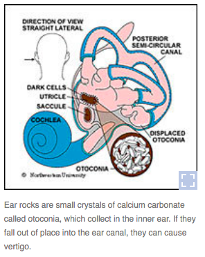
Inner ear crystals diagram. How to Cure Inner Ear Crystals | Healthfully Ear crystals (otolith or otoconia) are tiny calcium carbonate/calcite crystals embedded in the gelatinous otolithic membrane in the inner ear. The otolithic organs (the utricle and the saccule) are what enables you to discern which way is up even when your eyes are closed. Diagram Of Inner Ear Canal - Free Catalogs A to Z Inner Ear Diagram Photos and Premium High Res Pictures. Ear crystals: What they are and how they can cause. 4 hours ago Outer ear; Middle ear; Inner ear; View the diagrams below to learn more about the different sections of the ear and how we hear. Inner ear - Online Biology Notes The inner ear is also called as labyrinth because of its intricate structure of interconnecting chamber and passage. It consists of two main structural The sensory sport (macula) is present in both utriculus and sacculus. The macula consists of otolith membrane having otolith (small crystal of CaCO3) which... How To Balance Crystals In Your Ear The idea of crystals in your ear sounds very "new age," if not farfetched. But for those who experience sudden and unexplained dizziness, it is very real and can be quite frightening. Crystals (otoconia) are made of calcium, and they'll shift from either one or both of the otolith organs of the inner ear.
The Antatomy of Hearing and Balance Diagram of outer, middle, and inner ear. The outer ear is labeled in the figure and includes the ear canal. The middle ear includes the eardrum (tympanic The calcium crystals are the structures that ultimately stimulate the position hairs and provoke nerve impulses created by the position changes... Inner Ear Photos and Premium High Res Pictures - Getty Images Browse 1,560 inner ear stock photos and images available, or search for inner ear illustration or inner ear diagram to find more great stock photos and pictures. Illustration of hearing, journey of the sound wave in the ear. The sound wave is captured by the auricle, penetrates in the auditory canal, vibrates... Benign paroxysmal positional vertigo (BPPV) Exercise The method (for the left side) is performed as shown on the diagram. Stay in each of the supine (lying down) positions for 1 minute, and in the sitting upright position (top) for 1 minute. One cycle takes 4 minutes. The mirror image of this procedure is … Mayo Clinic Q and A: Dizziness Caused by Inner Ear Crystals Aug 06, 2016 · BPPV is a result of tiny crystals in your inner ear being out of place. The crystals make you sensitive to gravity and help you to keep your balance. Normally, a jelly-like membrane in your ear keeps the crystals where they belong. If the ear is damaged — often by a blow to the head — the crystals can shift to another part of the ear.
Magnetic Resonance Imaging Of Inner Ear MRI in inner ear pathology Congenital Inner ear anomalies. 6. Inner ear • Starts by 3rd week of fetal life and completed by 16th wks • The inner ear is derived from the ectoderm in the region of the hindbrain. • Benign Paroxysmal Positional Vertigo (BPPV) - VeDA BPPV is a mechanical problem in the inner ear. It occurs when some of the calcium carbonate crystals (otoconia) that are normally embedded in gel in the ... How the Inner Ear Balance System Works - Labyrinth... - YouTube How the Inner Ear Balance System Works - Labyrinth Semicircular Canals. Home Epley Maneuver | Johns Hopkins Medicine BPPV is caused by a problem in your inner ear. Your semicircular canals are found inside your ear. They detect motion and send this information to your brain. The utricle is a nearby part of the ear. It contains calcium crystals (canaliths) that help it detect movement.
Canalith repositioning procedure - Mayo Clinic 28 Aug 2020 — Vertigo usually comes from a problem with the part of the inner ear responsible for balance. BPPV occurs when tiny canalith particles (otoconia) ...
Generation of inner ear hair cells by direct lineage conversion of... (A) Diagram of the mouse inner ear shows the vestibular system (green) and the cochlea of the auditory system (red). Within the inner ear, expression of Atoh1, a bHLH class transcription factor (Lo et al., 1991; Ross et al., 2003) is both necessary and sufficient for the induction of sensory hair...
inner ear crystals Epley maneuver | Positional Vertigo: Get Medical... Crystals in my ears. This is how I spent the past 24 hours. I've been experiencing tiny dizzy spells for the past several months. Inner Ear 'Rock Slides' Lead To Vertigo. Tiny crystals, or ear rocks, in the inner ear help us to balance. But when these pebbles fall into the sensitive ear canal, they can...
Inner ear: bony and membranous labyrinths. Inner ear (labyrinthine): Semicircular canals, vestibule, cochlea (see the image below). Cross-section of the middle and inner ear. View Media Gallery. The ear is a multifaceted organ that connects Head reorientation relative to gravity induces movement of these crystals within the semicircular canal...
PDF Slide 1 | Twp parts of inner ear THE EAR: Diagram. 1. Pinna. Inner ear and Central auditory nervous system: When the stapes moves in and out of the oval window of the cochlea, it creates a fluid motion, hydrodynamic energy.
BPPV -- Benign Paroxysmal Positional Vertigo - Dizziness ... BPPV is caused by crystals dislodged from the utricle of the inner ear. In Benign Paroxysmal Positional Vertigo (BPPV) dizziness is thought to be due to debris ...
Inner Ear Crystals and BPPV These crystals are called Otoconia and are found in the Otolith organs, which are two pouches inside the vestibular system that are filled with fluid. When the head tilts, the crystals attached to the hairs move through the ear fluid and send nerve signals to the brain.
Why Loose Ear Crystals Make You Dizzy - Cleveland Clinic ... This crystal matrix serves as a reliable motion-sensing map — until crystals break free, drifting into one of the ear's three semicircular "balance" canals, and Why do loose crystals make you dizzy? Normally, the fluid in the semicircular canals and the small, direction-sensing cupula in your inner ear...
Adam J Calhoun on Twitter: "TIL we have...ear crystals?! And ...
Vestibular system: Anatomy, pathway and function | Kenhub 21.12.2021 · Vestibular system anatomy. The vestibular system is a somatosensory portion of the nervous system that provides us with the awareness of the spatial position of our head and body (proprioception) and self-motion (kinesthesia).). It is composed of central and peripheral portions. The peripheral portion of the vestibular system consists of the vestibular labyrinth, …
Ear crystals: What they are and how they can cause dizziness Jan 29, 2019 · When they are dislodged, the crystals float around in the fluid area of the balance branch of the inner ear, and you will start to feel off balance. The loose crystals will start to make people feel like they are spinning and the room is spinning around them. If you are 60 or older, you are more prone to having your ear crystals dislodge.
Eye - Wikipedia Eyes are organs of the visual system.They provide living organisms with vision, the ability to receive and process visual detail, as well as enabling several photo response functions that are independent of vision.Eyes detect light and convert it into electro-chemical impulses in neurons.In higher organisms, the eye is a complex optical system which collects light from the surrounding …
BeadDiagrams.com 10.2.2022 · To make a 7-inch bracelet (not including clasp), you will need about 23 crystals of each color (or 46 of one color). Each pair of crystals adds about 1/3-inch, if you want to make the bracelet longer (or shorter). More Photos and Demo Video. Click on the photos below (or the free beading pattern) to zoom in.
What Are the Symptoms of and Treatment for Crystals in the Inner Ear? Calcium chloride crystals dislodge inside the inner ear due to injuries involving sudden motion, aging and disease. In many patients with this condition The semicircular canals are fluid-filled structures in the inner ear that are responsible for balance. When loose calcium chloride crystals move into the...
Signs and Symptoms of Loose Inner Ear Crystals Ear crystals… You have probably never heard of them, but you have them. If you are experiencing vertigo or dizzy spells, they are most likely the ... Inside the utricle of the inner ear are small calcium crystals called otoconia. They are responsible for our sense of gravity and linear acceleration.
Cliff Pickover's RealityCarnival (09/22/17) Venn diagram: uppercase letter glyphs are shared by Greek, Latin & Cyrillic alphabets (09/21/17) Cortical atrophy in Alzheimer's Disease, associated with loss of gyri and sulci (09/20/17) Mathematics can be mystical and beautiful. Buddhabrot and Logistic Map (09/19/17) Real dandelion seed puffs preserved as they're about to disperse
Inner ear - human anatomy organs | INNER EAR DIAGRAM Inner ear anatomy. The inner ear is called a labyrinth.
Benign paroxysmal positional vertigo (BPPV) The crystals can become dislodged from their normal position for a number of reasons. These include a head injury or an infection of the inner ear. More commonly it happens for no reason. BPPV normally occurs in one ear but some people have it in both ears at the same time. Diagnosis Your description of your symptoms is helpful in diagnosing BPPV.
The inner ear - See the functions and parts of the inner ear The inner ear consist of the cochlea, the balance mechanism, the vestibular and auditory nerve. How does the inner ear function? Once the vibrations of the eardrum have been transmitted to the oval window, the sound waves continue their journey into the internal ear.
Inner ear - Wikipedia The inner ear (internal ear, auris interna) is the innermost part of the vertebrate ear. In vertebrates, the inner ear is mainly responsible for sound detection and balance. In mammals, it consists of the bony labyrinth...
Inner ear | Radiology Reference Article | Radiopaedia.org The inner ear refers to the bony labyrinth, the membranous labyrinth and their contents. It may also be referred to as the vestibulocochlear organ, supplied by the vestibulocochlear nerve (CN VIII). It is divided into three main parts: the cochl...
Human Ear Anatomy - Parts of Ear Structure, Diagram and ... Ear The internal (inner) ear is also called the labyrinth because of its complicated series of canals (Figure 4, 5 and 6). Structurally, it consists of two main divisions: an outer bony labyrinth that encloses an inner membranous labyrinth. It is like long balloons put inside a rigid tube. The bony labyrinth is a...
TV Archives - Hollywood.com Click to get the latest TV content. Sign up for your weekly dose of feel-good entertainment and movie content!
Structure and Functions of the Ear Explicated With Diagrams The inner ear houses the sensory organs that help in hearing and maintaining balance. The part of human ear involved in the function of hearing is the cochlea. These floating crystals cause serious balance and vertigo problems. How the Human Ear Functions.
The Delicate Balance of Ear Crystals | The Institute for Creation... So not only did ear crystals form in the wrong place, but they were misshapen and abnormally sized," according to co-author Kent Hill.2 So Not only are otoliths complex (being a crystalline arrangement of matter), but their timed and directed formation must result in the correct placement, shape, number...
Hearing - Inner Ear Development - Embryology The inner ear is derived from a pair of surface sensory placodes (otic placodes) that appear in human development during week 4 (GA week 6) in the head region lying behind the second pharyngeal arch. These otic placodes fold inwards forming initially a depression...
(PDF) The glycan keratan sulfate in inner ear crystals Mammalian otoconia of the inner ear vestibular apparatus are calcium carbonate-containing mineralized structures critical in maintaining balance and detecting linear acceleration. The mineral phase of otoconia is calcite, which coherently diffracts X-rays much like a single-crystal.
List of antibiotics - Wikipedia The following is a list of antibiotics.The highest division between antibiotics is bactericidal and bacteriostatic.Bactericidals kill bacteria directly, whereas bacteriostatics prevent them from dividing. However, these classifications are based on laboratory behavior. In practice, both treat a bacterial infection.
Canalith Repositioning Procedure (CRP) - Cleveland Clinic 22 Oct 2018 — Often the cause of vertigo is the displacement of small calcium carbonate crystals, or canaliths, within the inner ear.
Epley Maneuver to Treat BPPV Vertigo - YouTube 27 Sept 2014 — - Video demonstrates how the Epley maneuver is performed to treat POSTERIOR canal BPPV affecting the right ear ...
Inner ear: Anatomy | Kenhub The inner ear is embedded within the petrous part of the temporal bone, anterolateral to the posterior cranial fossa, with the medial wall of the middle ear These cells are covered by a gelatinous structure known as the otolithic or statoconial membrane and embedded in it are several small crystals known...
Do you or someone you know have misplaced BPPV Ear Crystals? Where are the ear crystals? Each inner ear, or vestibular system, has three semi-circular canals - the posterior, the anterior and the horizontal canal. Ear crystals live on a membrane in a nearby vestibular sensory organ called the utricle. All of the semi-circular canals share fluid with the utricle, so...
Inner Ear Diagram Stock Photos, Pictures & Royalty-Free ... Browse 176 inner ear diagram stock photos and images available, or search for human ear to find more great stock photos and pictures. Newest results. human ear. Human ear internal The ear is the organ that detects sound. It not only receives sound, but also aids in balance and body position.
The Inner Ear - Bony Labyrinth - Membranous... - TeachMeAnatomy The inner ear is located within the petrous part of the temporal bone. It lies between the middle ear and the internal acoustic meatus, which lie laterally and medially respectively. The inner ear has two main components - the bony labyrinth and membranous labyrinth.

0 Response to "37 Inner Ear Crystals Diagram"
Post a Comment