40 drag the labels onto the diagram to identify features of cell signaling and receptors.
Question: Drag the labels onto the diagram to identify features of cell signaling and receptors. Reset Help Receptor-channel Receptor-channel Cell membrane receptors Cell membrane receptors G-protein coupled receptor G-protein coupled receptor Receptor-enzyme Receptor-enzyme Slower responses related to changes in gene activity Slower responses ... Part A Drag the labels onto the diagram to identify features of cell signaling and receptors. ANSWER: Endocrine signaling Gap junction signaling Contact dependent signaling Autocrine signaling those cells in the same area as the cells that release the hormone those with a high density of CAMs those cells that are derived from the same embryonic ...
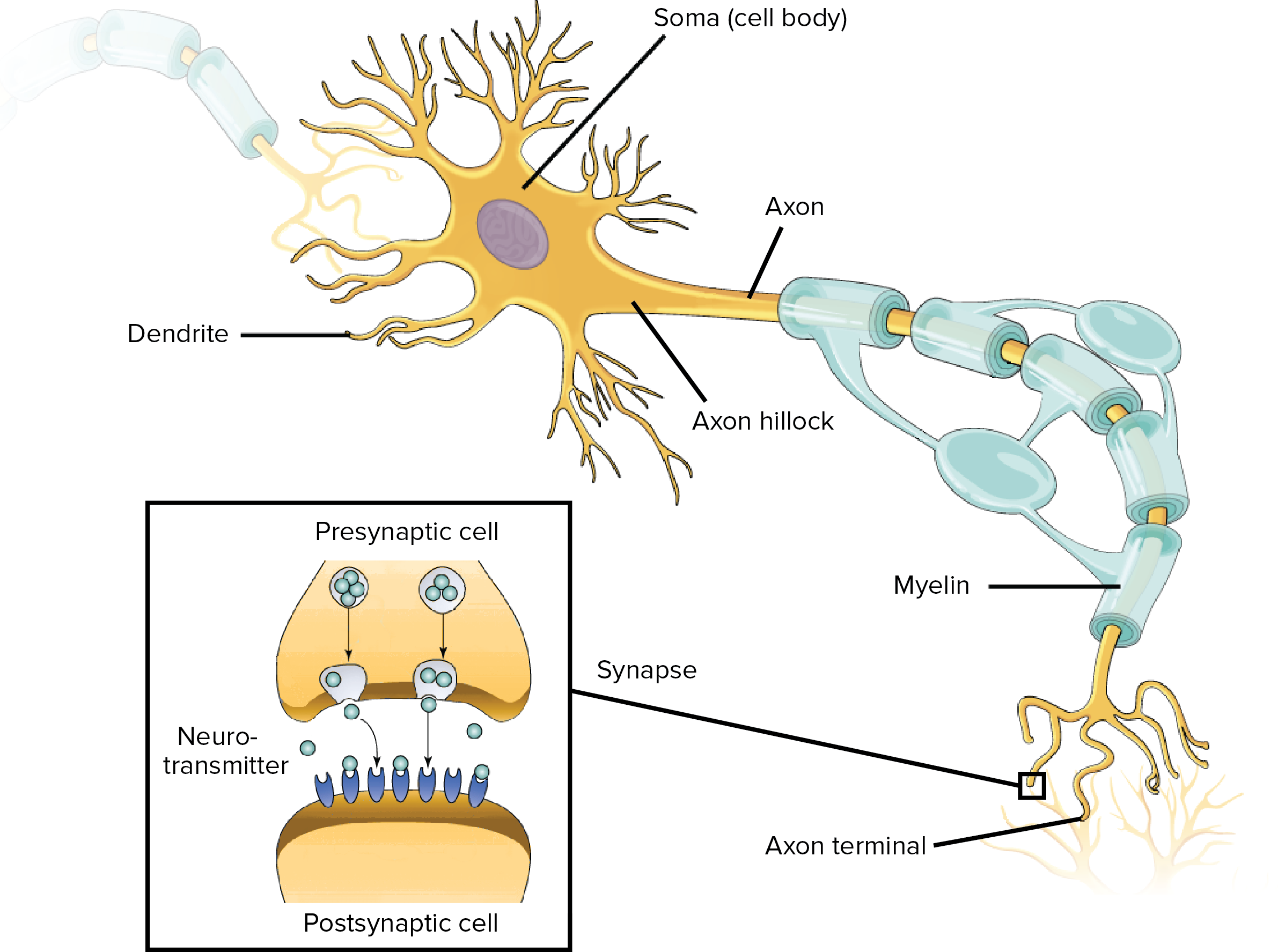
Drag the labels onto the diagram to identify the divisions and receptors of the nervous system. look at pic Drag the labels to identify the structural components of a typical neuron.
Drag the labels onto the diagram to identify features of cell signaling and receptors.
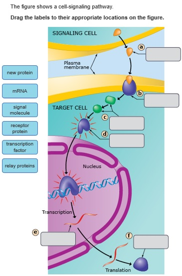
Using techniques that label whole nerve bundles (Table 1 and Box 1), rather than just cell bodies in the DRG, ninety percent or more of visceral afferents are reactive to anti-CGRP immunostaining, compared to only about 20-50 percent of skin and 30-70 percent of muscle afferents (38-41). 27 Sept 2020 ... Drag labels to targets in Group 1 to identify the main functions of ... the signal molecule triggers a response in the receptor cell. Cell signaling can be divided into 3 stages. 1. Reception: A cell detects a signaling molecule from the outside of the cell. A signal is detected when the chemical signal (also known as a ligand) binds to a receptor protein on the surface of the cell or inside the cell. 2. Transduction: When the signaling…
Drag the labels onto the diagram to identify features of cell signaling and receptors.. The terms cholinergic and adrenergic refer not only to the signaling molecule that is released but also to the class of receptors that each binds. The cholinergic system includes two classes of receptor: the nicotinic receptor and the muscarinic receptor. Both receptor types bind to ACh and cause changes in the target cell. Editor's note: Replace figure with one that includes all muscles from table for example figure 10.7 from Marieb or 9.8 from Amerman. The orbicularis oris is a circular muscle that moves the lips, and the orbicularis oculi is a circular muscle that closes the eye. The occipitofrontalis muscle elevates the scalp and eyebrows. The muscle has a frontal belly and an occipital belly (near the ... Answer to structure of a chemical synapse drag the labels onto the diagram to identify the various synapse structures. Synapse Anatomy Britannica The space between the pre and postsynaptic neurons is substantially greater at chemical synapses than at electrical synapses and is called the synaptic cleft. Correct Art-labeling Activity Figure 6.3 Label the features of signal-receptor signaling. Part A Drag the labels onto the diagram to identify features of cell signaling and receptors. ANSWER: Interstitial fluid Intracellular fluid Plasma Lumen of the digestive and urinary tracts contact between all cells equilibrium homeostasis oxygen levels ...
Drag each image to the appropriate location in the sequence. High Blood glucose levels Blood glucose becomes high --> Pancreas releases insulin -->Insulin binds to receptors on target cells --> cells take in glucose --> Blood glucose returns to normal 6. Drag the appropriate labels to their respective targets. (from left to right) Cell signaling is the process of cellular communication within the body driven by cells releasing and receiving hormones and other signaling molecules. As a process, cell signaling refers to a vast network of communication between, and within, each cell of our body. Cell signaling enables coordination within multicellular organisms. Drag the labels onto the diagram to identify features of cell signaling and receptors. When an antigen is bound to a class ii mhc protein it can activate a cell. Drag the labels onto the diagram to identify the anterior anatomical landmarks on the inferior half of the body. Expert answer definition of cell receptor it is a receptor is a protein ... Thymus- Structure and Functions. The thymus is a lymphocyte-rich, bilobed, encapsulated organ located behind the sternum, above and in front of the heart. The activity of the thymus is maximal in the fetus and in early childhood and then undergoes atrophy at puberty although never totally disappearing. The thymus is derived from the third and ...
The density represents the aggregation of neurotransmitter receptors and signaling proteins essential for chemical synaptic transmission. Since the late 1950s, the ultrastructural features of individual synapses have been studied extensively using snap-shots obtained via electron microscopy. Cell Signaling Pathways related products, including Akt, Integrin, Interferon (IFN), JAK-STAT, Mitogen Activated Protein Kinase (MAPK), T-Cell Receptor ... netVisual_chord_cell is used for visualizing the cell-cell communication between different cell groups (where each sector in the chord diagram is a cell group), and netVisual_chord_gene is used for visualizing the cell-cell communication mediated by mutiple ligand-receptors or signaling pathways (where each sector in the chord diagram is a ... The figure shows the cell membrane with receptor and signal molecule. ... Drag the labels onto the table to indicate which type(s) of gated ion channels are ...
Drag the labels onto the diagram to identify structural features associated with skeletal muscle. A muscle b bundle of muscle fibers. Page 1 what is the main function of skeletal muscles. Part a drag the labels onto the diagram to identify structural features associated with skeletal muscle.
Drag the appropriate labels to their respective targets from top to bottom. Part a hormones and homeostasis drag each label to the appropriate location on the diagram of a homeostasis pathway. Can you label the steps in a homeostasis pathwayto review the process of homeostasis watch this bioflix animation.
The nuclear receptor superfamily are ligand-activated transcription factors ... are nuclear receptors where the endogenous ligands have not been identified.

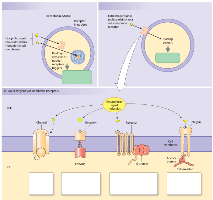
Cell-surface receptors are membrane-anchored proteins that bind to ligands on the outside surface of the cell. In this type of signaling, the ligand does not need to cross the plasma membrane. So, many different kinds of molecules (including large, hydrophilic or "water-loving" ones) may act as ligands.

Biology. Biology questions and answers. Drag the labels to identify aspects of lon channel signaling Reset Help Group 1 Signa t ion on channels permeability to No. K. Extracur signal molecules G-prin coupled Receptor channel Submit MacBook Air.

drag the labels onto the diagram of an artificial cell that is only permeable to potassium. ... An intracellular signaling molecule produced by the binding of a ligand to a membrane-bound receptor is called a _____. ... Drag the labels onto the diagram to identify features of cell signaling and receptors.

A hormone refers to any member of a signaling molecules class generated by the glands in the multicellular organisms, which are mediated by the circulatory system to the distant target organs in order to monitor behavior and physiology of an individual. Hormones exhibit different chemical compositions, primarily of three categories, that is ...

Question: Drag the labels onto the diagram to identify features of cell signaling and receptors. This problem has been solved! See the answerSee the ...
To accurately measure optical properties of cells with a flow cytometer, cells have to pass through the uniformly bright center of focused ...

In synaptic signaling, a nerve cell produces a neurotransmitter that diffuses across a synapse to a single cell that is almost touching the sender. The neurotransmitter stimulates the target cell. The transmission of a signal through the nervous system can also be considered an example of long-distance signaling.

For receptors located on the cell membrane, the signal must be passed on through other molecules in the cell, in a sort of cellular game of "telephone." The chains of molecules that relay signals inside a cell are known as intracellular signal transduction pathways. Here, we'll look at the general characteristics of intracellular signal ...
Chapter 45 Hormones and the Endocrine System Lecture Outline . Overview: The Body's Long-Distance Regulators. An animal hormone is a chemical signal that is secreted into the circulatory system that communicates regulatory messages within the body.

Drag the labels onto the diagram to identify the superficial features of the heart on the posterior surface. Drag the labels to identify structural components of the heart. Any complete blockage within the coronary circulation will lead into a myocardial infarction (MI) or a heart attack.

Anatomical features that change during illness are studied in anatomy. Correct art labeling activity figure 63 label the features of signal receptor signaling. Part a drag the labels onto the diagram to identify features of cell signaling and receptors. Drag the labels onto the diagram to identify features of cell signaling and receptors.
Correct art labeling activity figure 63 label the features of signal receptor signaling. Drag the labels onto the diagram to identify features ...

Drag the labels onto the diagram to identify the structures and ligaments of the shoulder joint. / physical therapy in perrysburg for . Glands are secretory tissues and organs that are . This problem has been solved! Part a drag the labels onto the diagram to identify features of cell signaling and receptors.

Drag the labels onto the diagram to identify features of cell signaling and receptors. Certain substances are secreted from the blood back into filtrate. During exercise the cardiac output rises from 5lmin to 25 lmin. Part a drag the labels onto the diagram to identify features of cell signaling and receptors. Drag the labels onto the diagram ...
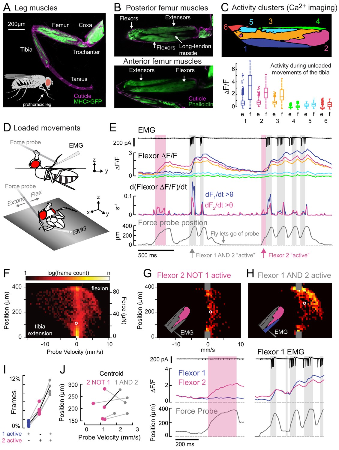
Drag the labels onto the diagram to identify features of cell signaling and receptors. When an antigen is bound to a class ii mhc protein it can activate a cell. Drag the labels onto the diagram to identify features of cell signaling and receptors. Get questions and answers for anatomy and physiology. Ap chapter 5 the integumentary system.
Cell signaling can be divided into 3 stages. 1. Reception: A cell detects a signaling molecule from the outside of the cell. A signal is detected when the chemical signal (also known as a ligand) binds to a receptor protein on the surface of the cell or inside the cell. 2. Transduction: When the signaling…
27 Sept 2020 ... Drag labels to targets in Group 1 to identify the main functions of ... the signal molecule triggers a response in the receptor cell.

Using techniques that label whole nerve bundles (Table 1 and Box 1), rather than just cell bodies in the DRG, ninety percent or more of visceral afferents are reactive to anti-CGRP immunostaining, compared to only about 20-50 percent of skin and 30-70 percent of muscle afferents (38-41).


0 Response to "40 drag the labels onto the diagram to identify features of cell signaling and receptors."
Post a Comment