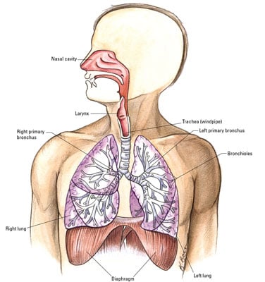
37 upper respiratory tract diagram
Main parts of the upper respiratory tract-Nose-Mouth-Pharynx-Epiglottis-Larynx -Trachea. First step of air entering the tract. Air enters through the nose. Nose function-Functions to protect the lower airway -Does this by warming air-Humidifying air-Filtering small particles before air enters the lungs. Students will be able to identify the parts of the respiratory system. Students will be able to describe the functions of each part of the respiratory system. Materials and preparation Poster board Computers Index cards (7 per student) Markers Respiratory System worksheet Your Respiratory System worksheet Notebook paper Are Your Lungs Healthy ...
Pharynx, epiglottis, trachea, larynx, tracheal cartilages, sinuses, and tonsils - anatomy and physiology of the upper respiratory tract. Find diagrams, notes...

Upper respiratory tract diagram
The respiratory system is situated in the thorax, and is responsible for gaseous exchange between the circulatory system and the outside world. Air is taken in via the upper airways (the nasal cavity, pharynx and larynx) through the lower airways (trachea, primary bronchi and bronchial tree) and into the small bronchioles and alveoli within the ... The respiratory tract in humans is made up of the following parts: External nostrils - For the intake of air.; Nasal chamber - which is lined with hair and mucus to filter the air from dust and dirt.; Pharynx - It is a passage behind the nasal chamber and serves as the common passageway for both air and food.; Larynx - Known as the soundbox as it houses the vocal chords, which are ... Start studying Upper Respiratory Tract Diagram. Learn vocabulary, terms, and more with flashcards, games, and other study tools.
Upper respiratory tract diagram. The lower respiratory tract includes the trachea, bronchi, and lungs (see Figure 1). Symptoms of upper respiratory tract infections include clear or colored discharge from the eyes or nose, coughing, sneezing, swelling of the mucous membranes around the eyes (conjunctivitis, see Figure 2), ulcers in the mouth, lethargy, and anorexia. 21st Century Family Survival Guide. Information we need to know about Corona viruses to understand this enemy. Upper Respiratory Tract Diagram. The respiratory system organs are separated into the upper and lower respiratory tracts. The upper respiratory tract includes the mouth, nose, nasal cavity, pharynx (windpipe and food pipe) and larynx or voice box. Each has a specific function to aid the flow of air into the body. In this article we will discuss about the structure of respiratory tract of humans with the help of suitable diagram. Also learn about its functions. i. Upper respiratory tract extends from the upper nares to the vocal cord. (Fig. 8.1) ii. Lower respiratory tract extends from the vocal cord to the alveoli.
Start studying Upper Respiratory Tract Diagram. Learn vocabulary, terms, and more with flashcards, games, and other study tools. Upper Respiratory Tract Structural and Functional Anatomy Nose and Nasal Cavity. The nostrils, the two round or oval holes below the external nose, are the primary entrance into the human respiratory system [5].Lying just after the nostrils are the two nasal cavities, lined with mucous membrane, and tiny hair-like projections called cilia [6].During inhalation, the air passes into the nasal ... Upper Respiratory Tract. Bones that form external nose. cartilage in external nose. skin over external nose supplied by. 3 parts of nasal cavity. frontal bone and frontal process of maxilla. Superior and inferior nasal cartilage, septal cartilage and sm…. external nasal, infratrochlear, Infraorbital nerves. The respiratory tract is divided into two sections, namely, upper and lower. The part above the voice box or larynx is upper respiratory tract and the one below it is lower respiratory tract. The respiratory tract is lined by respiratory mucosa or respiratory epithelium . The tract moistens and provides protection from pathogens and foreign bodies.
The upper respiratory tract is made up of the: Nose. Nasal cavity. Sinuses. Larynx. Trachea. The lower respiratory tract is made up of the: Lungs. Bronchi and bronchioles. Air sacs (alveoli) Lungs. The lungs take in oxygen. Your body's cells need oxygen to live and carry out their normal functions. The lungs also get rid of carbon dioxide, a ... The respiratory system is divided into an upper and lower respiratory tract. The upper tract comprises:. the nose and nasal cavity; the sinuses; the pharynx; the larynx The structures of your upper respiratory system are always working overtime—filtering air, expelling pollutants, and relying on other anatomy to remain healthy and intact. Let's take a look at some of the structures and jobs of the upper respiratory system. Take a look at the labeled diagram of the respiratory system above. As you can see, there are several structures to learn. Spend a few minutes reviewing the name and location of each one, then try testing your knowledge by filling in your own diagram of the respiratory system (unlabeled) using the PDF download below.
Upper respiratory tract: this part of the respiratory system extends from the nose to the trachea; sinuses ring the nose and upper jaw [NHLBI 2017.] Upper Respiratory Tract Diagram Frontal sinus Nostril Oral cavity Trachea. Title: B1162-002972-16_Sanofi_CSO_Allegra_Module1_Diagram_ADL02.indd
Upper Respiratory Tract Structural and Functional Anatomy Nose and Nasal Cavity. The nostrils, the two round or oval holes below the external nose, are the primary entrance into the human respiratory system [5].Lying just after the nostrils are the two nasal cavities, lined with mucous membrane, and tiny hair-like projections called cilia [6].During inhalation, the air passes into the nasal ...
The Dorsal Surface of the Tongue 10p Image Quiz. Compound Microscope and its Parts 16p Image Quiz. Skull of a Newborn--Diagram 10p Image Quiz. Photomicrograph of a C.S. of Skeletal Muscle 4p Image Quiz. Anatomy of the Composite Animal Cell--TEM 6p Image Quiz.
The respiratory tract is the path of air from the nose to the lungs. It is divided into two sections: Upper Respiratory Tract and the Lower Respiratory Tract. Included in the upper respiratory tract are the Nostrils, Nasal Cavities, Pharynx, Epiglottis, and the Larynx. The lower respiratory tract consists of the Trachea, Bronchi, Bronchioles,
Human respiratory system, the system in humans that takes up oxygen and expels carbon dioxide. The major organs of the respiratory system include the nose, pharynx, larynx, trachea, bronchi, lungs, and diaphragm. Learn about the anatomy and function of the respiratory system in this article.
Upper respiratory tract. Nasal cavity and nostrils: They mark the main entrance to the respiratory system as air passes through them into the next part of the airways. Paranasal sinuses: Air-filled spaces surrounding the nasal cavity.
The upper respiratory system, or upper respiratory tract, consists of the nose and nasal cavity, the pharynx, and the larynx. These structures allow us to breathe and speak. They warm and clean the air we inhale. mucous membranes lining upper respiratory structures trap some foreign particles,
The upper gastrointestinal tract consists of the mouth, pharynx, esophagus, stomach, and duodenum. The exact demarcation between the upper and lower tracts is the suspensory muscle of the duodenum.This differentiates the embryonic borders between the foregut and midgut, and is also the division commonly used by clinicians to describe gastrointestinal bleeding as being of either "upper" …
Upper Respiratory System. Create healthcare diagrams like this example called Upper Respiratory System in minutes with SmartDraw. SmartDraw includes 1000s of professional healthcare and anatomy chart templates that you can modify and make your own. 21/22 EXAMPLES. EDIT THIS EXAMPLE.
9.11.2021 · Upper respiratory tract. The upper respiratory tract refers to the parts of the respiratory system that lie outside the thorax, more specifically above the cricoid cartilage and vocal cords.It includes the nasal cavity, paranasal sinuses, pharynx and the superior portion of the larynx.Most of the upper respiratory tract is lined with the pseudostratified ciliated columnar epithelium, also ...
Upper Respiratory Tract Infections: Just as it sounds, upper respiratory tract infections occur in the upper respiratory system: nose/nostrils, nasal cavity, mouth, throat or pharynx, and larynx above the vocal folds. Examples of upper respiratory tract infections include sinusitis (also known as a sinus infection) and laryngitis (inflammation ...
10.11.2016 · Can upper respiratory tract infections be prevented? Prevention is difficult. Many germs (viruses) can cause an upper respiratory tract infection (URTI). Also, many viruses that cause URTIs are in the air, which you cannot avoid. However, the following are suggestions that may reduce the risk of catching a URTI or of passing one on, if you have ...
In humans and other mammals, the anatomy of a typical respiratory system is the respiratory tract.The tract is divided into an upper and a lower respiratory tract.The upper tract includes the nose, nasal cavities, sinuses, pharynx and the part of the larynx above the vocal folds.The lower tract (Fig. 2.) includes the lower part of the larynx, the trachea, bronchi, bronchioles and the alveoli.
A variety of viruses and bacteria can cause upper respiratory tract infections. These cause a variety of patient diseases including acute bronchitis, the common cold, influenza, and respiratory distress syndromes. Defining most of these patient diseases is difficult because the presentations connected with upper respiratory tract infections (URIs) commonly overlap and their causes are similar.
Upper respiratory tract: Composed of the nose, the pharynx, and the larynx, the organs of the upper respiratory tract are located outside the chest cavity. Nasal cavity: Inside the nose, the ...
As the names suggest, the upper respiratory tract includes everything above the vocal folds, furthermore, the lower respiratory tract includes everything below the vocal folds. Let us understand these tracts in further detail. The Upper Respiratory Tract. The upper respiratory tract starts with the sinuses and nasal cavity that are present in ...
The upper respiratory system, or upper respiratory tract, consists of the nose and nasal cavity, the pharynx, and the larynx. These structures allow us to breathe and speak. They warm and clean the air we inhale: mucous membranes lining upper respiratory structures trap some foreign particles, including smoke and other pollutants, before the ...
Start studying Upper Respiratory Tract Diagram. Learn vocabulary, terms, and more with flashcards, games, and other study tools.
The respiratory tract in humans is made up of the following parts: External nostrils - For the intake of air.; Nasal chamber - which is lined with hair and mucus to filter the air from dust and dirt.; Pharynx - It is a passage behind the nasal chamber and serves as the common passageway for both air and food.; Larynx - Known as the soundbox as it houses the vocal chords, which are ...
The respiratory system is situated in the thorax, and is responsible for gaseous exchange between the circulatory system and the outside world. Air is taken in via the upper airways (the nasal cavity, pharynx and larynx) through the lower airways (trachea, primary bronchi and bronchial tree) and into the small bronchioles and alveoli within the ...


0 Response to "37 upper respiratory tract diagram"
Post a Comment